Log in    RU  


Main page :: Forum :: Ultrasound cases - Sonograms, cine-loops etc. ::

Omphalocele and Tamm-Horsfall nephropathy


Tags: Soft tissues sonography, Urinary tract sonography, GE Logiq Book XP, Clinical report, Pediatric

   Posts
 
  Omphalocele and Tamm-Horsfall nephr...
#1

    A newborn at the neonatal pathology department of the Children's City Clinical Hospital with omphalocele, an ultrasound scan was prescribed with suspicion of hypoxic kidney damage and an omphalocele ultrasound scan.

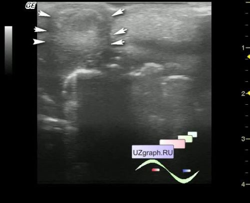
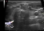
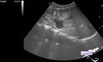
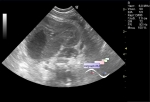
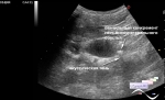
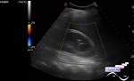
    On ultrasound of omphalocele it is visualized as a rounded structure, with contents of mixed echogenicity, without blood flow to the CFM, located in the anterior abdominal wall defect.

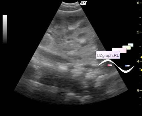
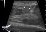
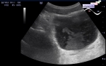
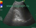
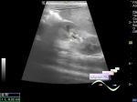
    On ultrasound of the kidneys, an increase in the echogenicity of the papillae of the renal pyramids is visualized - the syndrome of hyperechoic pyramids (Tamm-Horsfall nephropathy, by the name of the Tamm-Horsfall protein, which can cause hyperechoic renal pyramids in newborns and is considered a transient state, practically as a normal state if further on the follow-up ultrasound the indicated echo-pattern goes away!).


 Random 
Ultrasound cases - Double - IVH
    

Ultrasound cases - Bilateral popliteal cysts with legs
    

Scientific - Playlist Urinary tract sonography

Ultrasound cases - Kidney calcinate plus
    

Ultrasound cases - Diffuse heterogeneous liver
    

 Dictionary 
GS

TI-RADS

GA

TAV

PSV

EDV

PD

LMP

LVEDV

TAMEAN


 Calculators 
Volume (ellipse formula)

Teicholz systolic cardiac function

Gestational age by Ultrasound

TI-RADS

Myocardial mass


 Similar topics 
Ultrasound cases - Penetrating injury of the femur
    

Ultrasound cases - Cephalohematoma
    

Ultrasound cases - Urachus fistula
    

Ultrasound cases - Cranial mass
    

Ultrasound cases - Pilonidal coccyx cyst
    

Ultrasound cases - Palm fistula, in search of pencil lead
    

Ultrasound cases - Postoperative abdominal wall fistula
    

Ultrasound cases - Nucks cyst
    

Ultrasound cases - Paraproctitis
    

Ultrasound cases - Submandibular area lesion