Log in    RU  


Main page :: Forum :: Ultrasound cases - Sonograms, cine-loops etc. ::

Lesion of the temporal region


Tags: Soft tissues sonography, GE Logiq E, Clinical report, Pediatric

   Posts
 
  Lesion of the temporal region
#1

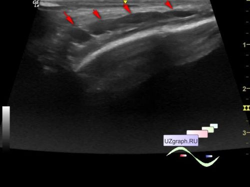
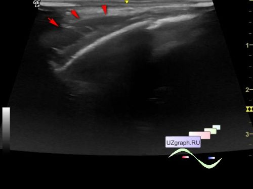
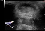
    Child 1 month old in the public clinic, after the parents applied with complaints of a slight swelling in the temporal region, the child was sent for an soft tissues ultrasound of the temporal region with suspicion of cephalohematoma.

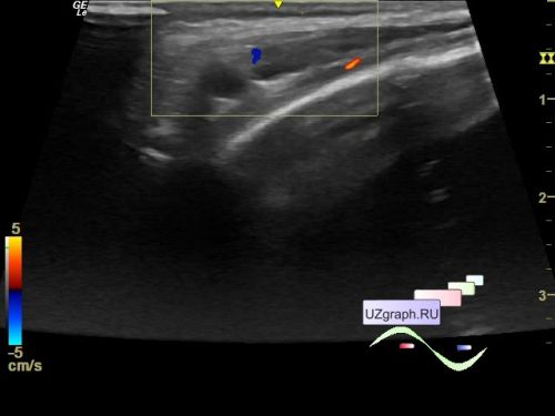
    In the projection of a lesion visible to the naked eye in the temporal region, an an-/hypoechoic lesion with septa or a group of lesions is visualized on ultrasound, the indicated area has a size of approximately 27 x 5 x 17 mm, at CFM the blood flow along the contour (differential diagnosis: lymphangioma, lymphadenitis, etc.)

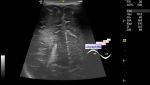
    The contralateral temporal region is shown at the end of the video for comparison.


 Random 
Ultrasound cases - Bicuspid aortic valve 2
    

Ultrasound cases - Lesion of the front wall of chest
    

Ultrasound cases - Extra flow in the RV
    

Ultrasound cases - Strangulated inguinal hernia with necrosis/hematoma
    

Ultrasound cases - Beads eggs
    

 Dictionary 
Aliasing doppler artifact

TV

FL

ESV

BSA

LMP

CRL

Qp-Qs

CFM

AC


 Calculators 
Volume (ellipse formula)

Pressure gradient

Gestational age by Ultrasound

Myocardial mass

Thyroid volume


 Similar topics 
Ultrasound cases - Penetrating injury of the femur
    

Ultrasound cases - Cephalohematoma
    

Ultrasound cases - Urachus fistula
    

Ultrasound cases - Cranial mass
    

Ultrasound cases - Pilonidal coccyx cyst
    

Ultrasound cases - Palm fistula, in search of pencil lead
    

Ultrasound cases - Postoperative abdominal wall fistula
    

Ultrasound cases - Nucks cyst
    

Ultrasound cases - Paraproctitis
    

Ultrasound cases - Submandibular area lesion