Log in    RU  


Main page :: Forum :: Ultrasound cases - Sonograms, cine-loops etc. ::

Subcutaneous lesion above the sternum


Tags: Soft tissues sonography, GE Logiq E, Clinical report, Pediatric

   Posts
 
  Subcutaneous lesion above the stern...
#1

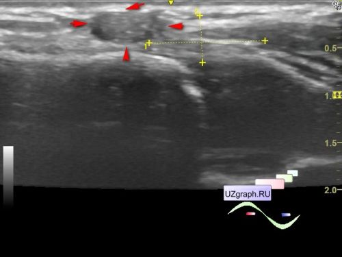
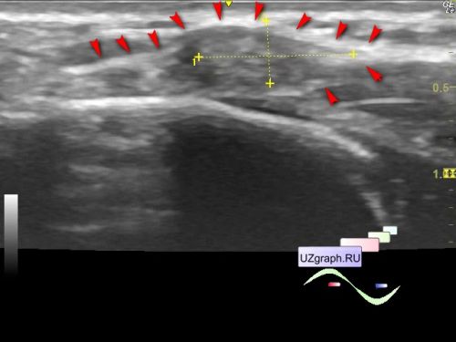
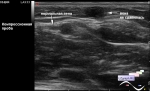
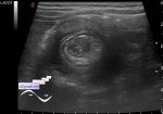
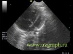
    A 9-year-old child was sent for a soft tissues ultrasound of the sternoclavicular joint to the public clinic after consulting of an orthopedist with complaints of a mass in the indicated projection.

    In the projection of complaints about a painful mass in the projection of the left sternoclavicular joint on ultrasound, a hypoechoic oval-shaped lesion with hyperechoic dot inclusions is visualized subcutaneously, with an "eye" sign - pushing apart the surrounding tissues with the formation of sharp corners and without clear boundaries in the lateral sections, up to 8 x 3 x 9 mm in size, at CFM without reliable signs of blood flow (differential diagnosis: schwannoma, etc.)


    external link


 Random 
Ultrasound cases - Tirads 5
    

Ultrasound cases - Flotation in the lateral ventricle
    

Ultrasound cases - Something on the neck
    

Ultrasound cases - Kidney's snowballs
    

Ultrasound cases - Lesion posterior to the thyroid right lobe
    

 Dictionary 
PW

TAMEAN

RAP

PD

LVOT

EDV

M-mode

GA

Gain

B-mode


 Calculators 
Myocardial mass

Gestational age by Ultrasound

Resistive index

Thyroid volume

TI-RADS


 Similar topics 
Ultrasound cases - Penetrating injury of the femur
    

Ultrasound cases - Cephalohematoma
    

Ultrasound cases - Urachus fistula
    

Ultrasound cases - Cranial mass
    

Ultrasound cases - Pilonidal coccyx cyst
    

Ultrasound cases - Palm fistula, in search of pencil lead
    

Ultrasound cases - Postoperative abdominal wall fistula
    

Ultrasound cases - Nucks cyst
    

Ultrasound cases - Paraproctitis
    

Ultrasound cases - Submandibular area lesion