Log in    RU  


Main page :: Forum :: Ultrasound cases - Sonograms, cine-loops etc. ::

Inter/intramuscular axillary mass


Tags: Soft tissues sonography, Esaote MyLab 70, Clinical report, Pediatric

   Posts
 
  Inter/intramuscular axillary mass
#1

    The parents of a 4-year-old child turned to the emergency department of the Children's City Clinical Hospital with complaints of painful mass in the axillary region, according to their words, the mass increased during long time.

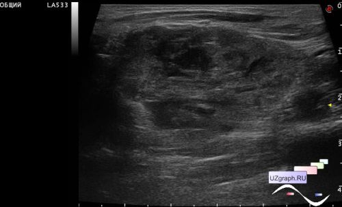
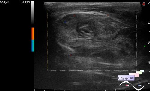
    At Ultrasound scan in the corresponding area visualized large inter- / intramuscular heterogeneous oval-shaped mass lesion intimately located with the bone, with a clear uneven contour, with a single blood flow signals at DPD.

    Diff. diagnosis: granular cell tumor, schwannoma, mts, etc.


 Random 
Ultrasound cases - Varicocele in a child
    

Ultrasound cases - Cystic dysplasia, hematuria
    

Ultrasound cases - Diaphragmatic hernia, intestine necrosis
    

Ultrasound cases - Hyperparathyroidism?


Ultrasound cases - Complication of a blood test from a vein
    

 Dictionary 
Freeze

LMP

MC

PRF

EFW

SV

LVEDD

FVI

LVPWd

EDV


 Calculators 
Body surface area

Type of an infant hip joint

Volume (ellipse formula)

Gestational age by Ultrasound

Myocardial mass


 Similar topics 
Ultrasound cases - Penetrating injury of the femur
    

Ultrasound cases - Cephalohematoma
    

Ultrasound cases - Urachus fistula
    

Ultrasound cases - Cranial mass
    

Ultrasound cases - Pilonidal coccyx cyst
    

Ultrasound cases - Palm fistula, in search of pencil lead
    

Ultrasound cases - Postoperative abdominal wall fistula
    

Ultrasound cases - Nucks cyst
    

Ultrasound cases - Paraproctitis
    

Ultrasound cases - Submandibular area lesion