Log in    RU  


Main page :: Forum :: Ultrasound cases - Sonograms, cine-loops etc. ::

Horseshoe kidney and Megaureter


Tags: Urinary tract sonography, Images, Video, Clinical report, GE Logiq Book XP, Pediatric

   Posts
 
  Horseshoe kidney and Megaureter
#1

    Infant with suspected L-shaped kidney by previous sonography exams addressed to follow-up US.

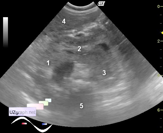
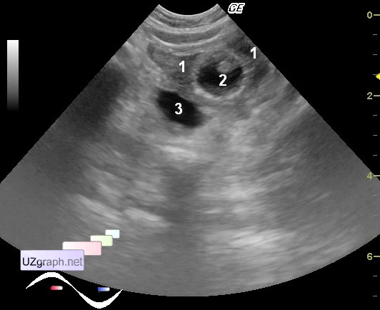
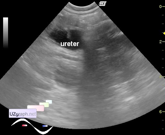
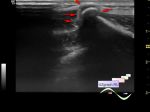
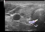
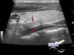
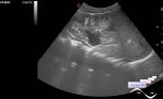
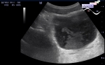
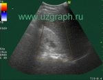
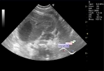
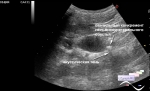
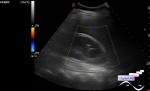
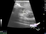
    At US left kidney didn' t visualized in the typical place from flank view. At scan thru anterior abdominal wall visualized left kidney(lobe) which communicate by isthmus with right kidney(attachment 1, see annotation below), long axises of kidneys are parallel, so it' s a variant of horseshoe kidney. Both renal pelvis are dilated. In the small pelvis right and down to empty bladder with Foley catheter visualized dilated ureter (megaureter - attachment 2, see annotation below) route of which is impossible to trace(attachment 3), in the left renal pelvis and right megaureter - visualized phenomenon of snow-storm(sludge?).

    Attachment 1:

    1 - right kidney(lobe)

    2 - isthmus

    3 - left kidney(lobe)

    4 - liver

    5 - vertebra

    Attachment 2:

    1 - bladder

    2 - Foley catheter

    3 - megaureter

    external link

:: attachments(4) ::

:: file 1 ::

:: file 2 ::

:: file 3 ::

:: file 4 ::

  16:24 09-06-2014
 
#2

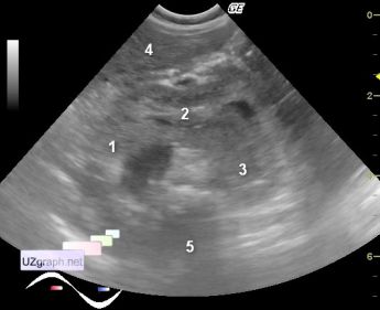
    Today I understood that didn't extract an image of dilated left renal pelvis from video and didn't explain one of structures in the first attachement to previous post - I'm very sorry(there is high humidity here in Moscow), and attaching missing images.

:: attachments(2) ::

:: file 1 ::

:: file 2 ::


 Random 
Ultrasound cases - Good vs Evil - femur
    

Ultrasound cases - Cholelithiasis 3
    

Ultrasound cases - Scapula exostosis
    

Ultrasound cases - Simple kidney cyst with calcification
    

Ultrasound cases - Torticollis 2
    

 Dictionary 
PRF

BSA

PG

PADP

ESV

HC

PASP

EDD

CO

GA


 Calculators 
Volume (ellipse formula)

Gestational age by Ultrasound

Body surface area

Qp:Qs

Pulmonary artery pressure


 Similar topics 
Ultrasound cases - Autosomal recessive polycystic kidney disease (ARPKD)
    

Ultrasound cases - Bladder sludge
    

Ultrasound cases - Bright kidneys
    

Ultrasound cases - Ileostoma stenosis and nephropathy
    

Ultrasound cases - Renal pelvis clot
    

Ultrasound cases - Renal pelvis valve-stone
    

Ultrasound cases - Condition after treatment of a kidney cyst
    

Ultrasound cases - Hydronephrotic transformation & polypous bladder wall
    

Ultrasound cases - Pyelitis, cystitis
    

Ultrasound cases - Lesion in the projection of the adrenal gland 2