Log in    RU  


Main page :: Forum :: Ultrasound cases - Sonograms, cine-loops etc. ::

Gluteal area oedema / panniculitis


Tags: Soft tissues sonography, Images, Video, Clinical report, Esaote MyLab 70, Pediatric

   Posts
 
  Gluteal area oedema / panniculitis
#1

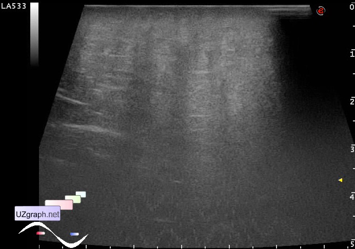
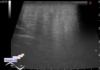
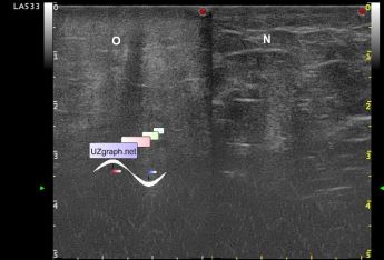
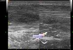
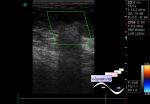
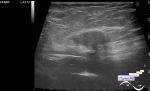
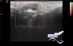
    A teenager with a suspected mass in the gluteal region was taken to the emergency department of the Children's City Clinical Hospital and urgently sent for an ultrasound scan.

    Visually, the buttocks look normal.

    At palpation in the area of complaints (the medial surface of the gluteal region) tissue has a diffusely increased density, crunches like wet snow.

    According to the patient, there were no injections and acne in this area before, this mass was found a few days ago.

    On ultrasound, a diffuse increase in the echogenicity of the subcutaneous fat, presumably edema / panniculitis, is visualized.

    Image legend: O - oedema, N - normal adipose tissue in symmetrical gluteal region.

:: attachments(3) ::

:: file 1 ::

:: file 2 ::

:: file 3 ::


 Random 
Ultrasound cases - Subdural hemorrhage 2
    

Ultrasound cases - Coronary fistula from LCA?
    

Ultrasound cases - Classic appendicular infiltrate
    

Ultrasound cases - Echogenic brain foci
    

Ultrasound cases - Zone of reduced echogenicity in the prostate
    

 Dictionary 
B-mode

EDV

SF

LVPWd

Gain

RAP

MC

PSV

EFW

AC


 Calculators 
Resistive index

Pulmonary artery pressure

Volume (ellipse formula)

Qp:Qs

Gestational age by Ultrasound


 Similar topics 
Ultrasound cases - Penetrating injury of the femur
    

Ultrasound cases - Cephalohematoma
    

Ultrasound cases - Urachus fistula
    

Ultrasound cases - Cranial mass
    

Ultrasound cases - Pilonidal coccyx cyst
    

Ultrasound cases - Palm fistula, in search of pencil lead
    

Ultrasound cases - Postoperative abdominal wall fistula
    

Ultrasound cases - Nucks cyst
    

Ultrasound cases - Paraproctitis
    

Ultrasound cases - Submandibular area lesion