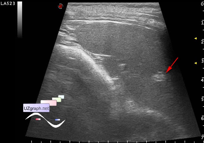
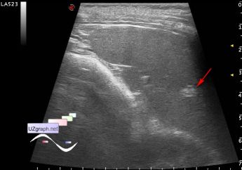
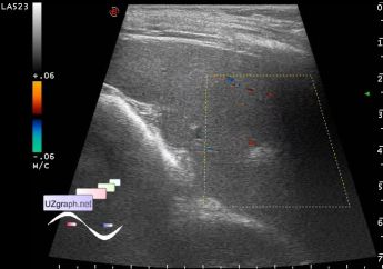
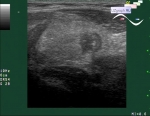
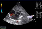
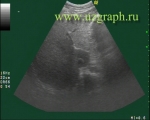
Log in    RU  


Main page :: Forum :: Ultrasound cases - Sonograms, cine-loops etc. ::

Spleen lesion


Tags: Abdomen sonography, Images, Video, Clinical report, Esaote MyLab 70, Pediatric

   Posts
 
  Spleen lesion
#1

    Child 10 years-old some time under control at one commercial center with accessory spleen and splenic hemangioma 5x5 mm.

    At current US at the border of the upper and middle thirds of the spleen visualized area of increased echogenicity in the course of vascularity(?).

    external link

:: attachments(3) ::

:: file 1 ::

:: file 2 ::

:: file 3 ::


 Random 
Ultrasound cases - Clinic is always right
    

Ultrasound cases - PDA 7yo
    

Ultrasound cases - Area with hyperechoic dot inclusions
    

Ultrasound cases - Cyst-like lesion in the back of the neck in infant
    

Ultrasound cases - Neck mass
    

 Dictionary 
EDV

IVSd

Freeze

TAV

LVPWd

MSD

LVESD

FL

SV

ESD


 Calculators 
Pulmonary artery pressure

Myocardial mass

Pressure gradient

Body surface area

TI-RADS


 Similar topics 
Ultrasound cases - Hirschsprung disease on ultrasound
    

Ultrasound cases - Intestinal Infiltrate
    

Ultrasound cases - Abdominal cryptorchidism
    

Ultrasound cases - Diffuse heterogeneous liver
    

Ultrasound cases - Postoperative abdominal wall fistula
    

Ultrasound cases - Stump appendicitis
    

Ultrasound cases - Liver scar after resection
    

Ultrasound cases - Subdiaphragmatic movable fluid collection (UFO)
    

Ultrasound cases - Lesion of the spleen or the case when it is easy to not notice
    

Ultrasound cases - I’m normal but others ...