Log in    RU  


Main page :: Forum :: Ultrasound cases - Sonograms, cine-loops etc. ::

Chronology of the coccyx's cyst


Tags: Soft tissues sonography, Images, Video, Clinical report, Esaote MyLab 70, Pediatric

   Posts
 
  Chronology of the coccyx's cyst
#1

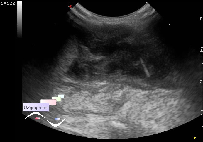
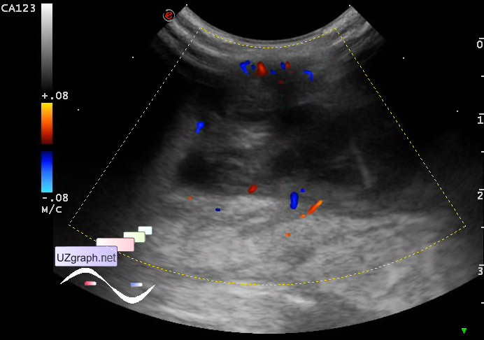
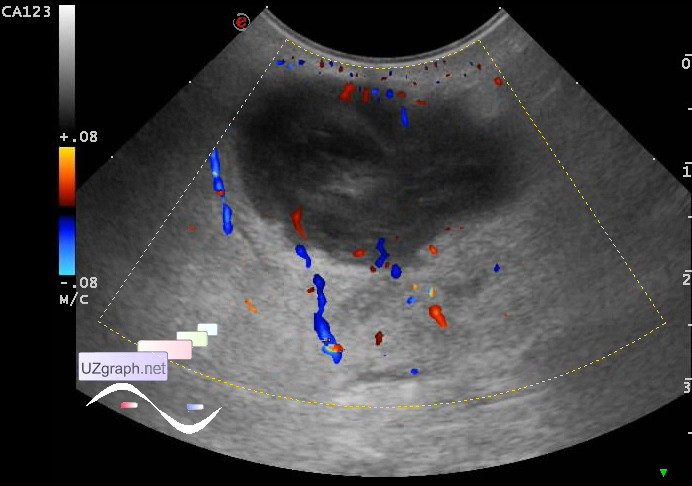
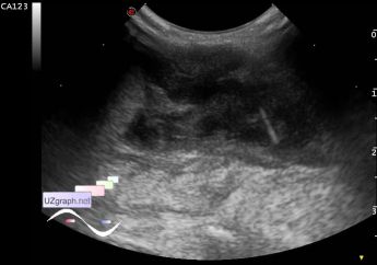
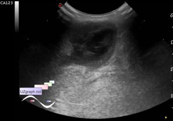
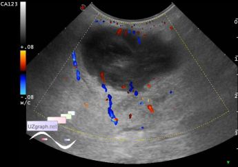
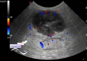
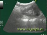
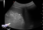
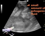
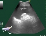
    A child 16 years old with complaints of pain in the coccyx when walking, can not sit. Already had raised this complaint in past, was diagnosed with a cyst of the coccyx and by the words of the patient, the contents of the cyst was evacuated thru syringe.

    Currently there is an area of skin redness in the coccyx area.

    On US in this projection is visualized irregular oval shaped heteroechoic lesion, type of complex cyst with a thick wall and heterogeneous content (hair?), size of 2x4cm, with increased blood flow at CFM, as well as in the surrounding tissues (inflammed pilonidal cyst?).

    external link

:: attachments(7) ::

:: file 1 ::

:: file 2 ::

:: file 3 ::

:: file 4 ::

:: file 5 ::

:: file 6 ::

:: file 7 ::


 Random 
Ultrasound cases - Cranial mass
    

Ultrasound cases - Hematoma of the gluteal region
    

Ultrasound cases - Periappendicular Abscess
    

Ultrasound cases - The effect of extreme
    

Ultrasound cases - Return of spermatocele
    

 Dictionary 
PADP

LVPWd

Qp-Qs

RAP

CW

Color M-mode

B-mode

Ved

PD

HR


 Calculators 
Resistive index

Pulmonary artery pressure

Body surface area

Qp:Qs

Volume (ellipse formula)


 Similar topics 
Ultrasound cases - Penetrating injury of the femur
    

Ultrasound cases - Cephalohematoma
    

Ultrasound cases - Urachus fistula
    

Ultrasound cases - Cranial mass
    

Ultrasound cases - Pilonidal coccyx cyst
    

Ultrasound cases - Palm fistula, in search of pencil lead
    

Ultrasound cases - Postoperative abdominal wall fistula
    

Ultrasound cases - Nucks cyst
    

Ultrasound cases - Paraproctitis
    

Ultrasound cases - Submandibular area lesion