Log in    RU  


Main page :: Forum :: Ultrasound cases - Sonograms, cine-loops etc. ::

Both ovaries teratomas


Tags: Female pelvis sonography, GE Logiq 400 MD, Images, Video, Clinical report, Pediatric

   Posts
 
  Both ovaries teratomas
#1

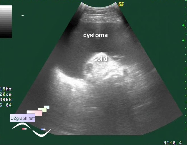
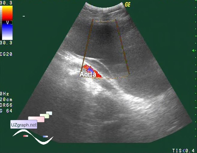
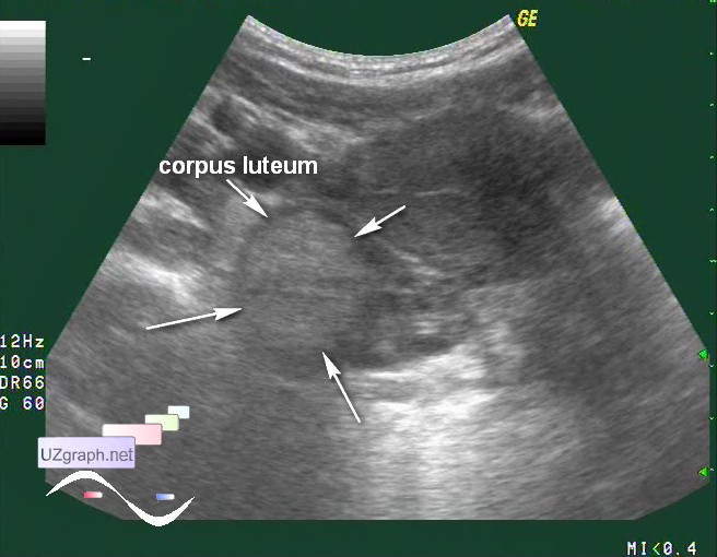
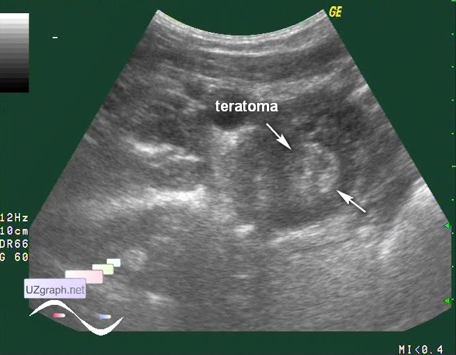
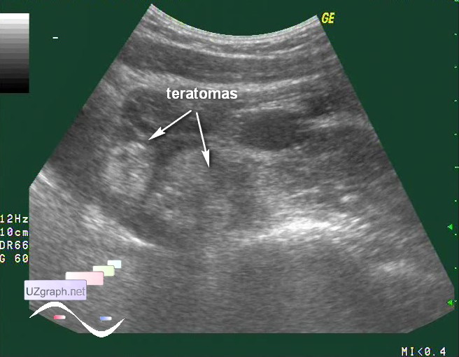
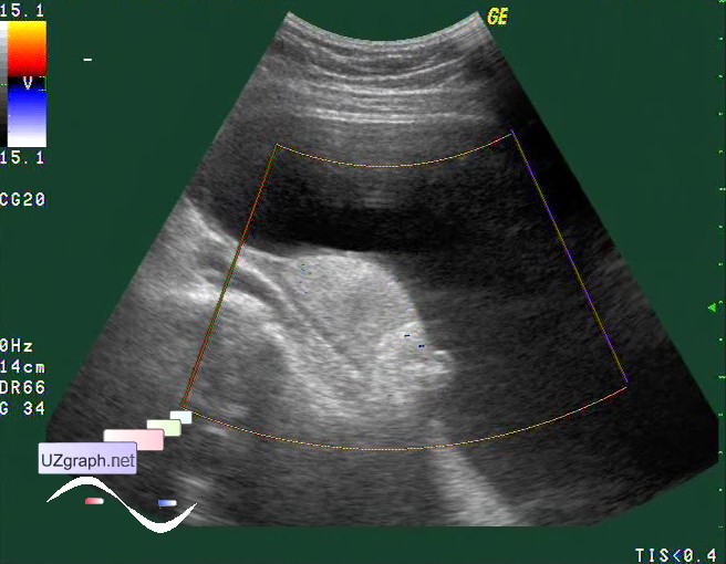
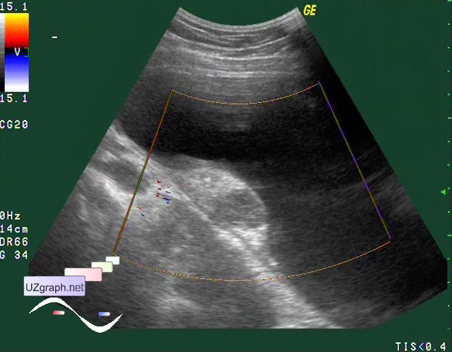
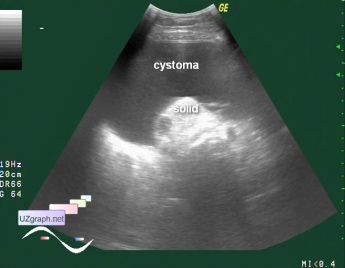
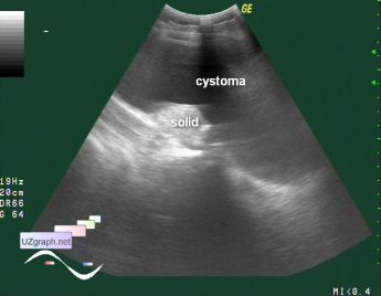
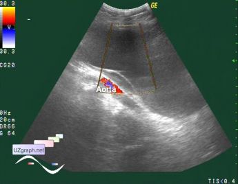
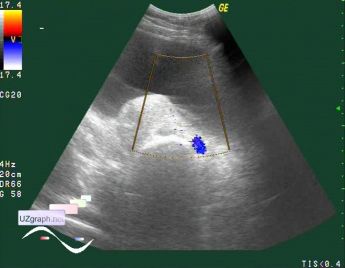
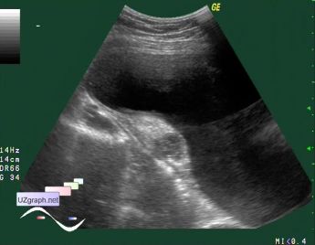
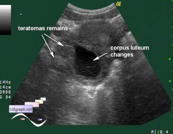
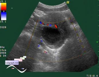
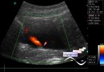
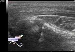
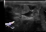
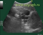
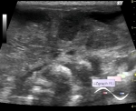
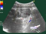
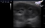
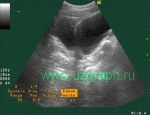
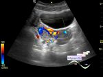
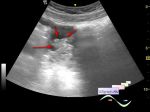
    A girl 16 years old went to the doctor complaining of tension feeling in the abdomen, is aimed at abdominal ultrasound, for unknown reason decided to drink water before the study - not need for abdominal US.

    Visually abdomen has abnormal shape - prominent low abdomen.

    At US practically the entire abdomen (mostly right lateral channel) is filled by giant cyst with some solid component below (cystoma), which is only a couple of cm does not reach the liver.

    In the right ovary was visualized hyperechoic (like solid) mass or masses.

    In a survey during the study it appears that there was a delay in menstrual cycle for about six months, with further questioning it turns out that 2-3 days ago was menstruating.

    The control ultrasound after 2 weeks, M-echo corresponds to second phase of MC, cystoma with no changes, solid mass in the right ovary turned into a cyst, most likely, the corpus luteum.

    Patient was aimed to oncologist and has operative intervention in oncocenter:

    " Clinical diagnosis: Left giant size ovarian teratoma. Teratomas of the right ovary. Functional cyst of the right ovary with hemorrhage into the cavity.

    Surgery: Laparoscopy. Enucleation of the left ovary teratoma. Resection of the left ovary. Enucleation of the right ovary two teratomas. Enucleation of the right ovary cyst."

    external link

:: attachments(16) ::

:: file 1 ::

:: file 2 ::

:: file 3 ::

:: file 4 ::

:: file 5 ::

:: file 6 ::

:: file 7 ::

:: file 8 ::

:: file 9 ::

:: file 10 ::

:: file 11 ::

:: file 12 ::

:: file 13 ::

:: file 14 ::

:: file 15 ::

:: file 16 ::


 Random 
Ultrasound cases - Portal hypertension
    

Ultrasound cases - Megaureter, ureteral valve-stone
    

Ultrasound cases - Suprasellar lession
    

Ultrasound cases - 99 Luftballons
    

Ultrasound cases - Anechogenic inclusion in the posterior cranial fossa
    

 Dictionary 
LVESV

VTI

HR

RAP

TI-RADS

GS

FVI

RVOT

DPD

Color M-mode


 Calculators 
Resistive index

Body surface area

Volume (ellipse formula)

Estimated fetal weight by Ultrasound

Pressure gradient


 Similar topics 
Ultrasound cases - Nucks cyst
    

Ultrasound cases - Hydrosalpinx (saktosalpinx)
    

Ultrasound cases - Portal vein air in the newborn
    

Ultrasound cases - Ovarian teratoma
    

Ultrasound cases - Inguinal hernia with an ovary
    

Ultrasound cases - Giant cyst of the right lateral canal
    

Ultrasound cases - Right ovary changes
    

Ultrasound cases - Ovarian apoplexy 3
    

Ultrasound cases - Hemorrhagic ovarian cyst
    

Ultrasound cases - Patchy uterus