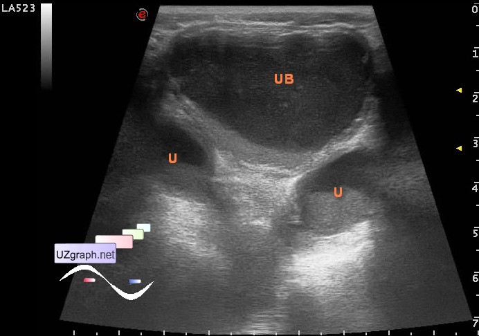
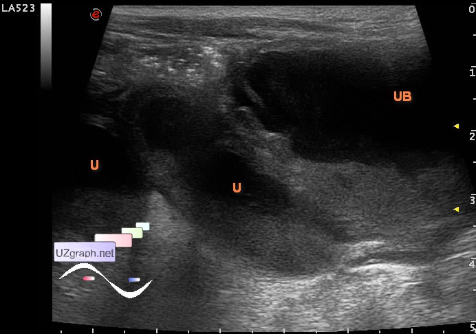
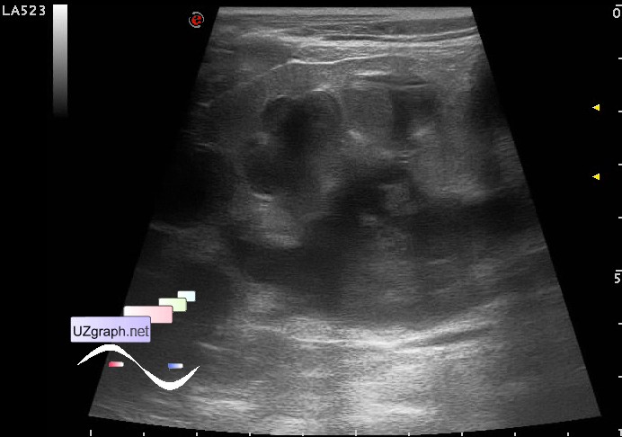
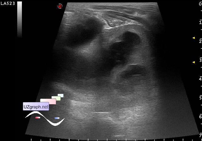
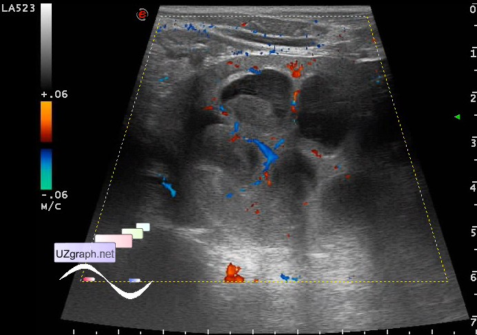
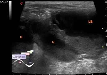
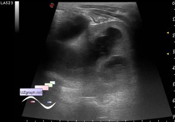
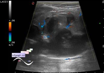
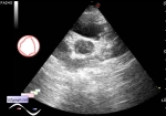
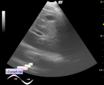
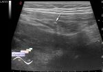
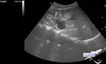
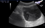
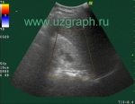
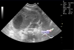
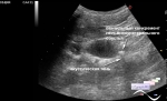
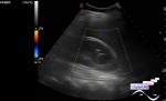
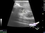
| Child 2 years old with a primary diagnosis of dysfunction of the VPS (ventriculo-peritoneal shunt) from the neurosurgery department, presumably with comorbidity, not walking, cachexia (skin and bones), skin color like white marble, in the lumbar region of the spine visualized area of raying through the transparent skin of the red color soft tissue with regular oval shape, head with a pronounced predominance of the cerebral part of the skull (2 times or more, " alien" head) to the face part. Directed to ultrasound of the abdomen and kidney due to changes specific for the inflammation in blood test, in particular a high ESR(erythrocyte sedimentation rate). According to the words of accompanying person is under control in another hospital with diagnosis of hydronephrosis / megaureter, currently in urinalysis a lot of bacteria, white blood cells. At ultrasound: kidneys (size very approximately, do not fit into the aperture of available probes due to too close placing of kidney with respect to the probe, have no any water-caps so was used the panorama scan with a known error of measurement): right 76x43mm parenchyma 6mm; left 61x41mm parenchyma 6mm; CMD(corticomedullary differentiation) is not visualized. Urinary tract(UT) is dilated: a calyces to 11mm on both sides, the pelvises to 28mm on both sides, in the UT lumen there is a sludge. The ureters are dilated all the way to 17 mm on both sides, in the lumen there is a lot of sludge. The bladder is filled, in the lumen there is a lot of sludge. Resume: Bilateral hydronephrosis / megaureter, possible UT infection. external link | |