Log in    RU  


Main page :: Forum :: Ultrasound cases - Sonograms, cine-loops etc. ::

Kidney calcinate plus


Tags: Urinary tract sonography, Thyroid gland sonography, GE Vivid 3, Images, Video, Clinical report, Adult

   Posts
 
  Kidney calcinate plus
#1

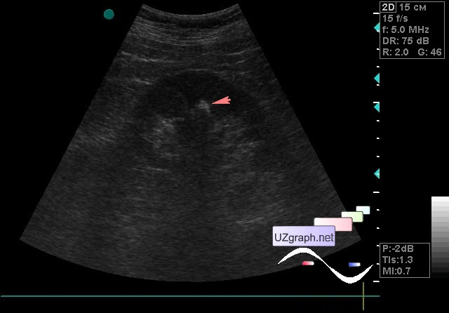
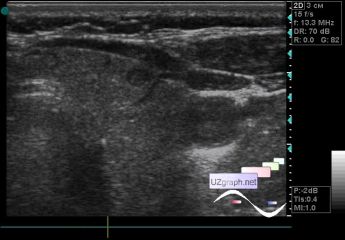
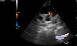
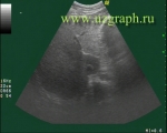
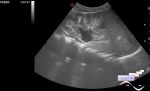
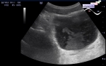
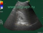
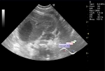
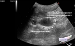
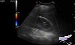
    The patient 75 years old came to the US of kidney because at US previously identified stones.

    No stones were detected on the current ultrasound but a hyperechoic inclusion was visualized with an acoustic shadow in the parenchyma up to 8 mm(calcinate? calcificated papilla of the pyramid? others?)

    Also, at the request of the patient, the thyroid gland was examined, as she said, previously was found a node there.

    On ultrasound in the left lobe a hypoechoic lesion of horizontal orientation up to 4 mm in size is visualized, at CFM without blood flow (colloid node? other?)

:: attachments(3) ::

:: file 1 ::

:: file 2 ::

:: file 3 ::


 Random 
Ultrasound cases - Flotation in the lateral ventricle
    

Ultrasound cases - Laparoscopic appendectomy, postoperative complications
    

Ultrasound cases - Complex cyst above the testicle
    

Ultrasound cases - Pediatric Tirads 5
    

Ultrasound cases - Suspicion of gastritis or renal colic
    

 Dictionary 
FNA

DPD

TAV

Gain

TV

IVSd

EDV

PG

BPD

LVPWd


 Calculators 
Thyroid volume

Pressure gradient

Gestational age by Ultrasound

Type of an infant hip joint

Body surface area


 Similar topics 
Ultrasound cases - Autosomal recessive polycystic kidney disease (ARPKD)
    

Ultrasound cases - Bladder sludge
    

Ultrasound cases - Bright kidneys
    

Ultrasound cases - Ileostoma stenosis and nephropathy
    

Ultrasound cases - Renal pelvis clot
    

Ultrasound cases - Renal pelvis valve-stone
    

Ultrasound cases - Condition after treatment of a kidney cyst
    

Ultrasound cases - Hydronephrotic transformation & polypous bladder wall
    

Ultrasound cases - Pyelitis, cystitis
    

Ultrasound cases - Lesion in the projection of the adrenal gland 2