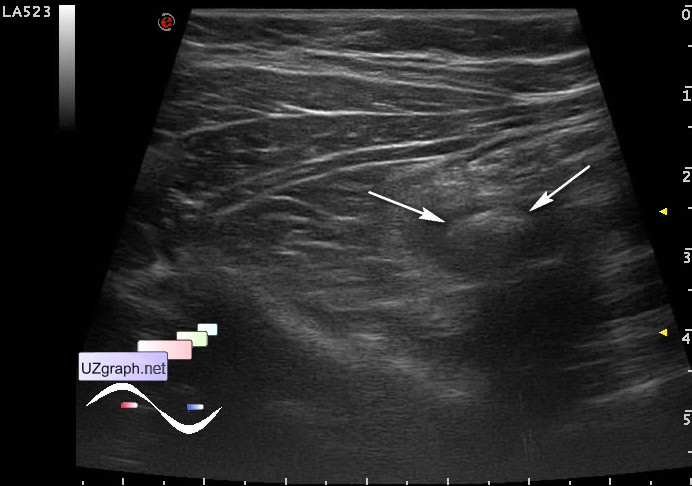
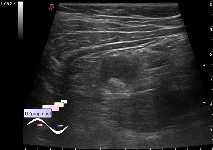
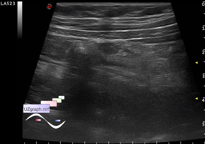
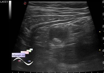
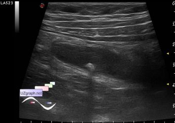
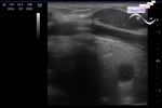
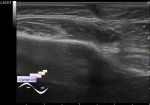
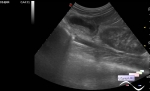
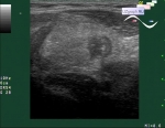
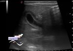
Log in    RU  


Main page :: Forum :: Ultrasound cases - Sonograms, cine-loops etc. ::

Appendicoliths


Tags: Gastrointestinal sonography, Images, Video, Clinical report, Esaote MyLab 70, Pediatric

   Posts
 
  Appendicoliths
#1

    A child 10 years old with suspected appendicitis.

    In the right side of the abdomen is visualised tubular structure of 10-14mm in diameter, the lumen mostly anechoic with hyperechoic inclusions, one in the middle to 8 mm and one at the bottom to 11mm (appendicoliths? acute appendicitis?)

    external link

:: attachments(5) ::

:: file 1 ::

:: file 2 ::

:: file 3 ::

:: file 4 ::

:: file 5 ::


 Random 
Ultrasound cases - Immunization schedule
    

Ultrasound cases - Condition after treatment of a kidney cyst
    

Ultrasound cases - Enlargement of the appendix
    

Ultrasound cases - Tirads 5
    

Ultrasound cases - Hydronephrosis of half of horseshoe kidney
    

 Dictionary 
PSV

ESV

MSD

HC

LVESV

IVC

VTI

IVSd

ESD

RVOT


 Calculators 
Pulmonary artery pressure

Qp:Qs

Body surface area

Myocardial mass

Type of an infant hip joint


 Similar topics 
Ultrasound cases - Hirschsprung disease on ultrasound
    

Ultrasound cases - Intestinal Infiltrate
    

Ultrasound cases - Subhepatic appendicitis and omentitis
    

Ultrasound cases - Diffuse heterogeneous liver
    

Ultrasound cases - Stump appendicitis
    

Ultrasound cases - Ileostoma stenosis and nephropathy
    

Ultrasound cases - Periappendicular infiltrate
    

Ultrasound cases - Intestinal obstruction, GB wall thickening
    

Ultrasound cases - Ileotiphlitis and mesadenitis
    

Ultrasound cases - Acute Appendicitis, periappendicular infiltrate