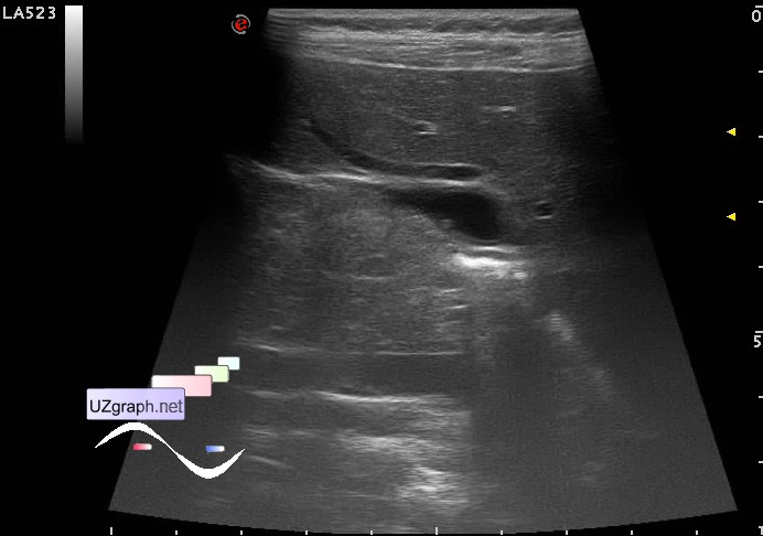
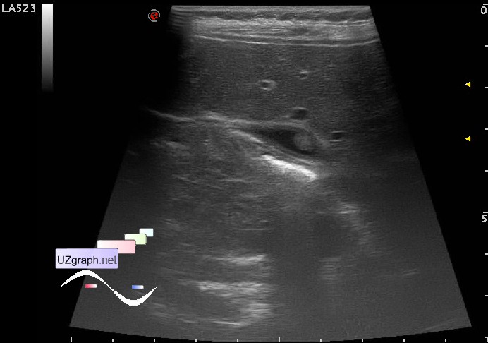
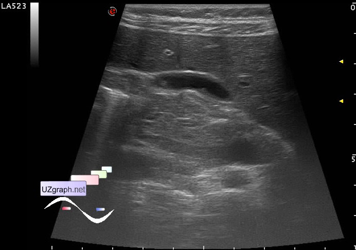
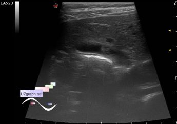
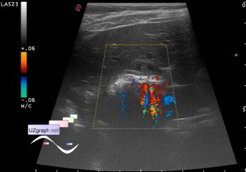
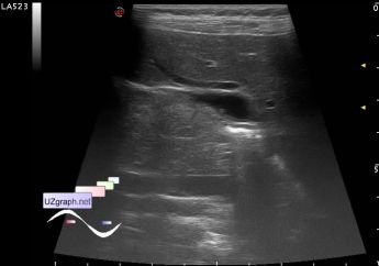
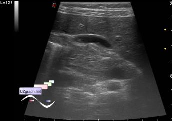
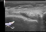
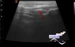
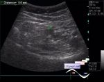
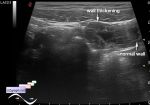
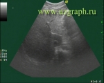
Log in    RU  


Main page :: Forum :: Ultrasound cases - Sonograms, cine-loops etc. ::

Gallbladder polyps


Tags: Abdomen sonography, Esaote MyLab 70, Images, Video, Clinical report, Pediatric

   Posts
 
  Gallbladder polyps
#1

    A child 14 years old with suspected appendicitis.

    At US in the projection of the gallbladder lumen visualized multiple (4 or more) parietal hypoechoic lesion of different sizes, the largest in the body, size of about 5 mm, without blood flow at CFM (GB polyposis / cholesterol polyps?).

    external link

:: attachments(6) ::

:: file 1 ::

:: file 2 ::

:: file 3 ::

:: file 4 ::

:: file 5 ::

:: file 6 ::


 Random 
Ultrasound cases - Portal vein air in the newborn
    

Ultrasound cases - Lesion in the right upper quadrant
    

Ultrasound cases - Extreme spleen
    

Ultrasound cases - Riedels lobe vs diaphragmatic hernia
    

Ultrasound cases - Cholelithiasis plus 2
    

 Dictionary 
VTI

EF

LVESV

PW

M-mode

EDD

SF

CFM

LVEDV

HC


 Calculators 
Resistive index

Estimated fetal weight by Ultrasound

Gestational age by Ultrasound

Qp:Qs

Type of an infant hip joint


 Similar topics 
Ultrasound cases - Hirschsprung disease on ultrasound
    

Ultrasound cases - Intestinal Infiltrate
    

Ultrasound cases - Abdominal cryptorchidism
    

Ultrasound cases - Diffuse heterogeneous liver
    

Ultrasound cases - Postoperative abdominal wall fistula
    

Ultrasound cases - Stump appendicitis
    

Ultrasound cases - Liver scar after resection
    

Ultrasound cases - Subdiaphragmatic movable fluid collection (UFO)
    

Ultrasound cases - Lesion of the spleen or the case when it is easy to not notice
    

Ultrasound cases - I’m normal but others ...