Log in    RU  


Main page :: Forum :: Ultrasound cases - Sonograms, cine-loops etc. ::

Neuroblastoma. Diffuse infiltration of liver


Tags: Urinary tract sonography, Abdomen sonography, Aloka Alpha 6, Images, Video, Clinical report, Pediatric

   Posts
 
  Neuroblastoma. Diffuse infiltration...
#1

    Child 2 months old in intensive care unit with a diagnosis of a right adrenal gland tumor (neuroblastoma).

    Appointed US due to the progressive enlargement of the abdomen (fluid?).

    Consulted by surgeon, it suggested that it' s a swollen bowel loops.

    Visually abdomen sharply enlarged.

    At US almost all the abdominal cavity filled by the liver which is visualized even in the pelvis, right lobe antero-posterior size in the right upper quadrant (!) 8cm, length about 15 cm (probe in the pelvis).

    In the projection of the lower pole of the right kidney visualized echogenic lesion (with slightly higher echogenicity than hepatic), with anechoic inclusions, size of 4x3 cm, with blood flow at CFM; left kidney is normal.

    In the lower left quadrant of the abdomen / pelvis visualized (subhepatic) bowel loops and a small amount of free fluid.

    external link

:: attachments(9) ::

:: file 1 ::

:: file 2 ::

:: file 3 ::

:: file 4 ::

:: file 5 ::

:: file 6 ::

:: file 7 ::

:: file 8 ::

:: file 9 ::

  10:16 25-04-2016
 
#2

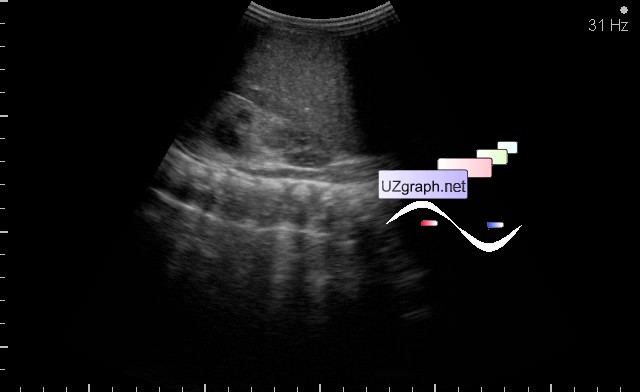
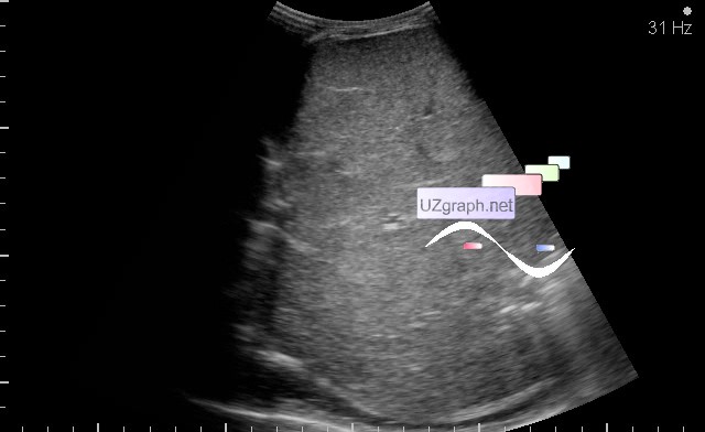
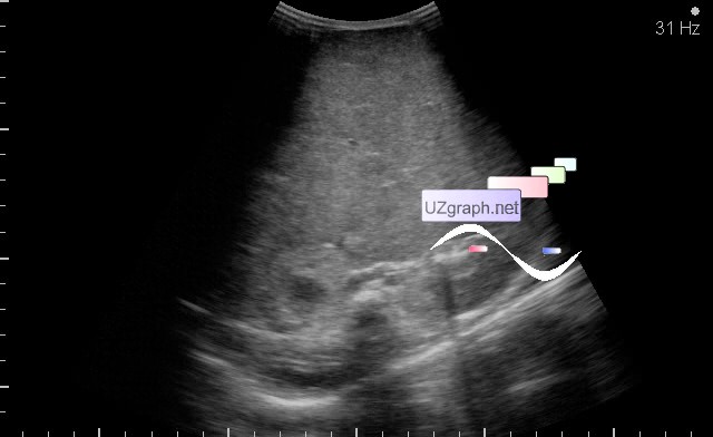
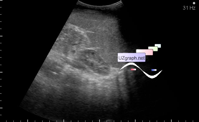
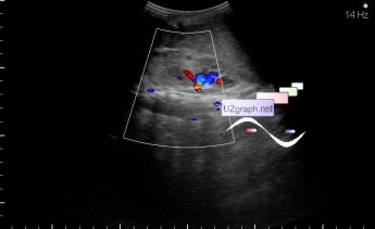
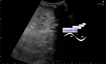
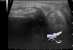
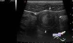
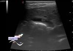
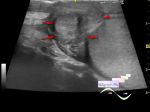
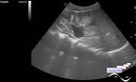
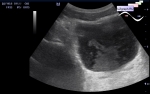
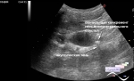
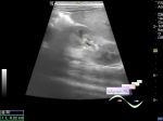
    To the question of normal adrenal gland imaging, as it's often done in some medical centers, measured the soft tissue above the upper pole of the kidney and everyone is happy.

    In this case, the adrenal lesion is localized in the projection of the lower renal pole because adrenal gland, in practice, are not always localized in the projection of the upper pole of kidney, and usually not visualized on ultrasound.

    While taking into account the localization in this case could be suspected nephroblastoma (Wilms' tumor) but as I spoke before the case was verified.

    external link

    "Wilms Tumor Imaging

    ...

    Abdominal neuroblastomas usually develop in the retroperitoneum. Most arise from the adrenal gland and displace the kidney inferomedially. In rare cases, a neuroblastoma may mimic a Wilms tumor, arising from tissues in the kidney or invading the kidney. To make diagnosis complicated, rare neuroblastomas possess other features more typical of Wilms tumor than of intrarenal neuroblastomas."

    As in the present case, the tumor site is not a typical arrangement of the adrenal gland, kidney does not displaced inferomedially but vice versa ...


 Random 
Ultrasound cases - Osgood-Schlatter disease, tendinitis
    

Admin room - New flash-components

Ultrasound cases - Electrode in the right heart after installing of heart pacemaker
    

Ultrasound cases - Diffuse-focal changes in the liver, thick left kidney
    

Ultrasound cases - Megaureter and cystic kidney dysplasia
    

 Dictionary 
VTI

BSA

EFW

TAV

M-mode

TVUS

FNA

LVESV

EDV

Anatomical M-mode


 Calculators 
Resistive index

Pulmonary artery pressure

Volume (ellipse formula)

Body surface area

Myocardial mass


 Similar topics 
Ultrasound cases - Ileostoma stenosis and nephropathy
    

Ultrasound cases - Renal pelvis clot
    

Ultrasound cases - Renal pelvis valve-stone
    

Ultrasound cases - Condition after treatment of a kidney cyst
    

Ultrasound cases - Hydronephrotic transformation & polypous bladder wall
    

Ultrasound cases - Pyelitis, cystitis
    

Ultrasound cases - Lesion in the projection of the adrenal gland 2
    

Ultrasound cases - Hyperechoic renal sinus with acoustic shadow
    

Ultrasound cases - Lesions in the projection of the adrenal glands
    

Ultrasound cases - Parietal bladder lesion