Log in    RU  


Main page :: Forum :: Ultrasound cases - Sonograms, cine-loops etc. ::

Torticollis - infiltrate


Tags: Soft tissues sonography, Musculoskeletal sonography, Esaote MyLab 70, Images, Video, Clinical report, Pediatric

   Posts
 
  Torticollis - infiltrate
#1

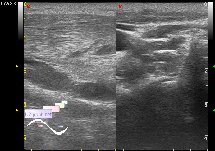
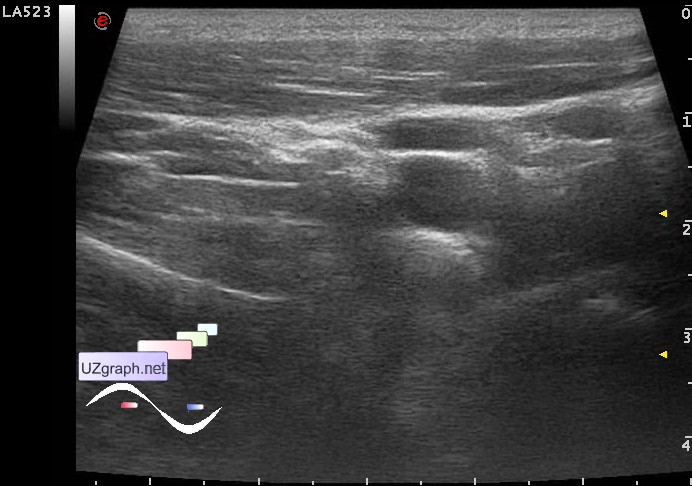
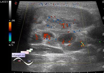
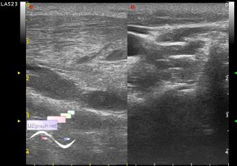
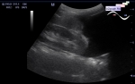
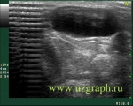
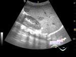
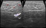
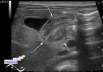
    A child 16 years old with suspected abscessed cervical lymphadenitis.

    In the side area of the neck there is severe (more than 2 times as compared with the other side - Fig. 3-5) thickening of the sternocleidomastoid muscle, in the posterior segments of muscle there is increased blood flow at CFM, posterior margin of muscle is unclear. To the posterior margin of the muscle adjoin a group of LN (about 10) maximum of 2 cm, in the area of an unclear muscule margin the LN also lose margin clarity, some of them in this area with the blood flow at CFM, some without, also near there is a isoechoic area, type of sludge contents (pus?), this area totally is about 3x4x1,5 cm(infiltrate with the abscess? etc.?).

    external link

:: attachments(6) ::

:: file 1 ::

:: file 2 ::

:: file 3 ::

:: file 4 ::

:: file 5 ::

:: file 6 ::


 Random 
On-call room - USB-killer
    

Ultrasound cases - Hydronephrotic transformation & polypous bladder wall
    

Ultrasound cases - Thyroid lesions
    

Ultrasound cases - Horseshoe kidney and Megaureter
    

Ultrasound cases - Cyst of the base of the brain
    

 Dictionary 
CRL

IVC

LMP

LVESD

SF

Aliasing doppler artifact

IVSd

ESD

LVEDV

M-mode


 Calculators 
Pulmonary artery pressure

Pressure gradient

TI-RADS

Resistive index

Qp:Qs


 Similar topics 
Ultrasound cases - Penetrating injury of the femur
    

Ultrasound cases - Cephalohematoma
    

Ultrasound cases - Urachus fistula
    

Ultrasound cases - Cranial mass
    

Ultrasound cases - Pilonidal coccyx cyst
    

Ultrasound cases - Palm fistula, in search of pencil lead
    

Ultrasound cases - Postoperative abdominal wall fistula
    

Ultrasound cases - Nucks cyst
    

Ultrasound cases - Paraproctitis
    

Ultrasound cases - Submandibular area lesion