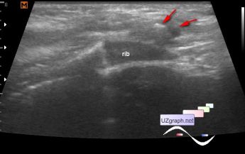
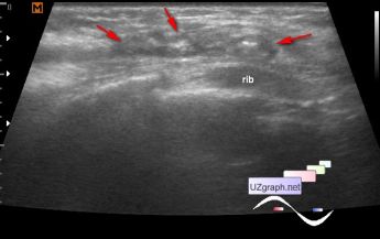
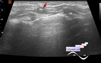
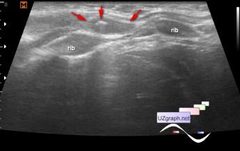
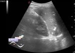
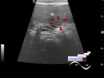
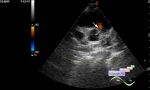
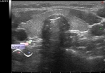
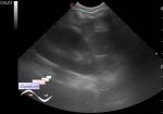
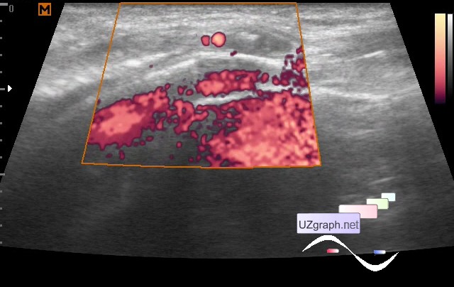
Lesion of the front wall of chest
Tags: Soft tissues sonography, Medison Sonoace R7, Images, Video, Clinical report, Pediatric
| Posts | |||
| Lesion of the front wall of chest | #1 |
| |||||
:: file 1 ::
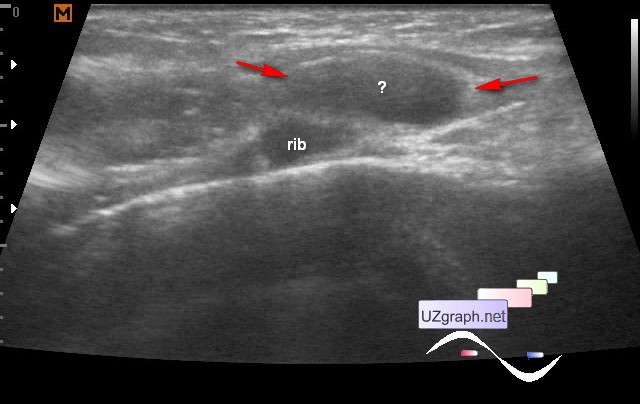 :: file 2 ::
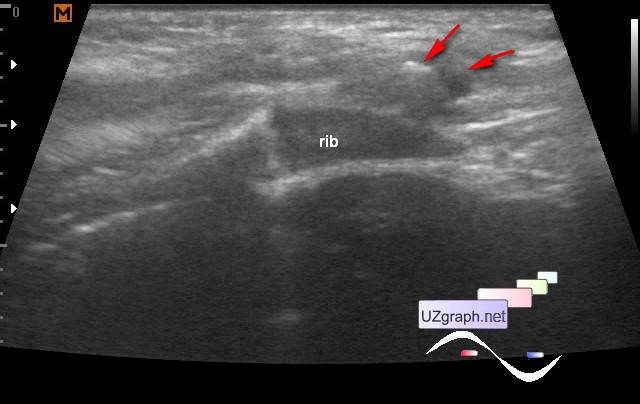 :: file 3 ::
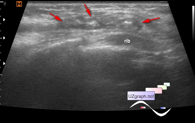 :: file 4 ::
 :: file 5 ::
 :: file 6 ::
 :: file 7 :: | |||||