Log in    RU  


Main page :: Forum :: Ultrasound cases - Sonograms, cine-loops etc. ::

Uterine myoma(fibroid)


Tags: Female pelvis sonography, GE Vivid 3, Images, Video, Clinical report, Adult

   Posts
 
  Uterine myoma(fibroid)
#1

    A 19-year-old patient came to the university public clinic for the abdomen ultrasound with complaints of abdominal pain.

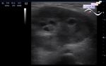
    On abdomen ultrasound without features.

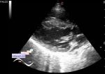
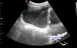
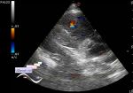
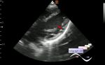
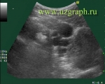
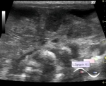
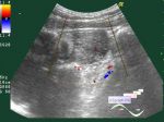
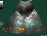
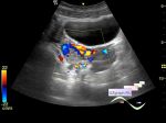
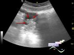
    In the projection of the bottom of the uterus, a hypoechoic lesion(perhaps several but one for sure) deforming the uterus contour is visualized, up to 2x1 cm in size, on the CFM the blood flow along the contour (myoma/ fibroid?).

    According to the patient, previously on ultrasound had been suspected a myoma but then after consilium it was decided that there was no fibroids after all.

    external link

:: attachments(3) ::

:: file 1 ::

:: file 2 ::

:: file 3 ::


 Random 
Ultrasound cases - Thickening and heterogeneity of the intercostal space soft tissues
    

Ultrasound cases - Lumbar pain
    

Ultrasound cases - Twinkling artifact
    

Ultrasound cases - Lesions of mammary glands
    

 Dictionary 
RVOT

EDV

IVSd

PSV

M-mode

ESD

LVOT

SF

Anatomical M-mode

PA


 Calculators 
Qp:Qs

Thyroid volume

Estimated fetal weight by Ultrasound

Volume (ellipse formula)

Gestational age by Ultrasound


 Similar topics 
Ultrasound cases - Nucks cyst
    

Ultrasound cases - Hydrosalpinx (saktosalpinx)
    

Ultrasound cases - Portal vein air in the newborn
    

Ultrasound cases - Ovarian teratoma
    

Ultrasound cases - Inguinal hernia with an ovary
    

Ultrasound cases - Giant cyst of the right lateral canal
    

Ultrasound cases - Right ovary changes
    

Ultrasound cases - Ovarian apoplexy 3
    

Ultrasound cases - Hemorrhagic ovarian cyst
    

Ultrasound cases - Patchy uterus