Log in    RU  


Main page :: Forum :: Ultrasound cases - Sonograms, cine-loops etc. ::

Diaphragmatic hernia, intestine necrosis


Tags: Chest sonography, Abdomen sonography, Gastrointestinal sonography, Cardiac sonography(Echocardiography), GE Logiq Book XP, Images, Video, Clinical report, Pediatric

   Posts
 
  Diaphragmatic hernia, intestine nec...
#1

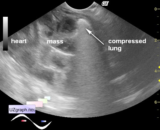
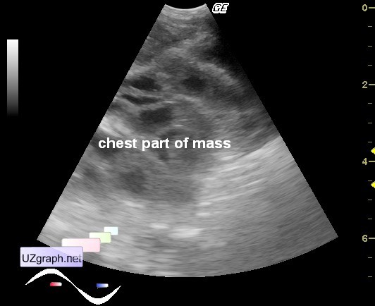
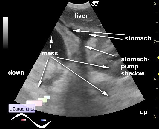
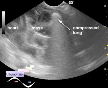
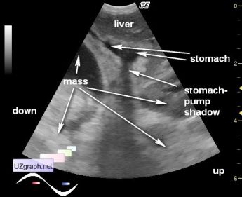
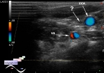
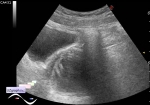
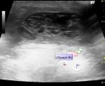
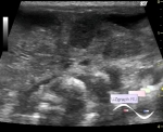
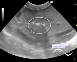
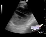
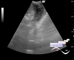
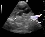
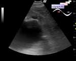
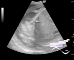
    Newborn with diaphragmatic hernia in the urgent therapy block. Surgeon palpated some mass in the abdomen and called the ultrasound.

    At sonography all abdomen and most of left chest consists of heterogeneous (cystic-solid) mass. At CFM abdominal part of the mass has well blood supply and no blood supply visualized in the chest part of the mass. About 5 ml of free fluid with echogenic dot particles in the subhepatic space.

    For verification of the abdominal part of the mass under ultrasound control was instilled fluid thru the stomach-pump - it is defined as a thin tubular structure compressed by the mass to the liver.

    Then was instilled fluid per rectum - fluid visualized as the content in the tubular structure in the mid-abdomen(center of mass). But some hollow structures weren't look like an intestine, they consisted of filiform structures which moved in time with heart beat and respiration movements.

    Surgeon made a suspicion that mass is an intestine with oedema of mesentery and omentum. I told honestly that I can't differentiate this mass from neo-process. In my memory was several cases of carcinomatosis with an identical sonographic picture.

    Child urgently taken into the operative intervention room. Intraoperative diagnosis - subtotal intestine necrosis. Surgeon tell that it's a very rare case...

    external link

:: attachments(10) ::

:: file 1 ::

:: file 2 ::

:: file 3 ::

:: file 4 ::

:: file 5 ::

:: file 6 ::

:: file 7 ::

:: file 8 ::

:: file 9 ::

:: file 10 ::


 Random 
Ultrasound cases - Daughters-mothers
    

Ultrasound cases - Few weeks after appendectomy
    

Ultrasound cases - Condition after diaphragmatic hernia
    

Ultrasound cases - Umbilical hernia in infant
    

Ultrasound cases - Inflamed mammary cyst(mastitis)
    

 Dictionary 
MC

AC

TVUS

HR

EDV

PW

LMP

HC

PD

Color M-mode


 Calculators 
Thyroid volume

TI-RADS

Volume (ellipse formula)

Body surface area

Qp:Qs


 Similar topics 
Ultrasound cases - Chylopericardium, lung atelectasis, cardiomegaly, ascites
    

Ultrasound cases - Diaphragmatic hernia, condition after surgery
    

Ultrasound cases - Hydrothorax
    

Ultrasound cases - One and a half kilograms of happiness
    

Ultrasound cases - Mirror artifact vs collapsed lung
    

Ultrasound cases - Persistent thymus or not
    

Ultrasound cases - Air bronchogram
    

Ultrasound cases - Postoperative hydrothorax with sludge
    

Ultrasound cases - Pneumonia and hydrothorax on the left
    

Ultrasound cases - Diaphragmatic hernia, ectopic spleen & air...