Log in    RU  


Main page :: Forum :: Ultrasound cases - Sonograms, cine-loops etc. ::

Hemorrhagic ovarian cyst & fluid sac in uterus cervix


Tags: Female pelvis sonography, Images, Video, Clinical report, Esaote MyLab 70, Pediatric

   Posts
 
  Hemorrhagic ovarian cyst & fluid sa...
#1

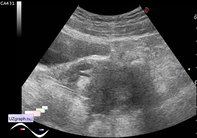
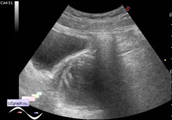
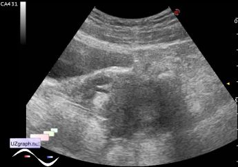
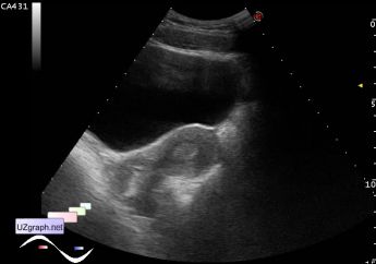
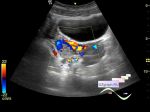
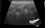
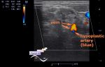
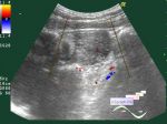
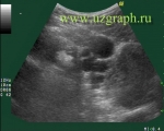
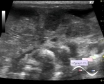
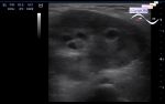
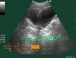
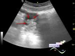
    Teenager with clinically suspected acute appendicitis was addressed to urgent abdomen ultrasound.

    At ultrasound an vermiform appendix did not visualized due to intestinal content which is blocks ultrasound.

    Under the not fully distended bladder was partially visualize some large cystic mass(about 4 cm) and fluid inclusion in the projection of the uterus cervix.

    Was recommended female pelvis sonography after the right preparation(filling of the bladder).

    At the female pelvis sonography(end of menstrual cycle) after some hours well defined cystic mass 4x5cm with thick wall in the right iliac area(projection of complaints on pain) - differential diagnosis: hemorrhagic cyst of the right ovary, etc. In the cervix of the uterus visualized some anechoic inclusion (diff. diagnosis: EGS, fluid in the cervical canal, etc.). At the uterus fundus visualized two canals(diff. diagnosis: duplication of the uterus, etc.).

    external link

:: attachments(11) ::

:: file 1 ::

:: file 2 ::

:: file 3 ::

:: file 4 ::

:: file 5 ::

:: file 6 ::

:: file 7 ::

:: file 8 ::

:: file 9 ::

:: file 10 ::

:: file 11 ::


 Random 
Ultrasound cases - Cyst of spermatic cord
    

Ultrasound cases - Infiltrate/ phlegmon of the wrist
    

Ultrasound cases - Parathyroid lesions
    

Ultrasound cases - Accessory thymic lobe
    

Ultrasound cases - Lesion in pancreas head
    

 Dictionary 
LMP

IVC

BSA

CW

CO

PW

LVESV

TAV

Ved

FVI


 Calculators 
Estimated fetal weight by Ultrasound

TI-RADS

Thyroid volume

Pulmonary artery pressure

Gestational age by Ultrasound


 Similar topics 
Ultrasound cases - Right ovary changes
    

Ultrasound cases - Ovarian apoplexy 3
    

Ultrasound cases - Hemorrhagic ovarian cyst
    

Ultrasound cases - Patchy uterus
    

Ultrasound cases - Fibroid and nabothian cyst
    

Ultrasound cases - Small pelvis lesion, presumably hydrocolpos
    

Ultrasound cases - Something in the female pelvis
    

Ultrasound cases - Uterine myoma(fibroid)
    

Ultrasound cases - Giant mass in the female pelvis
    

Ultrasound cases - Gallbladder, free fluid, etc.