Log in    RU  


Main page :: Forum :: Ultrasound cases - Sonograms, cine-loops etc. ::

Clinically suspected appendicitis


Tags: Female pelvis sonography, Images, Video, Clinical report, Esaote MyLab 70, Pediatric

   Posts
 
  Clinically suspected appendicitis
#1

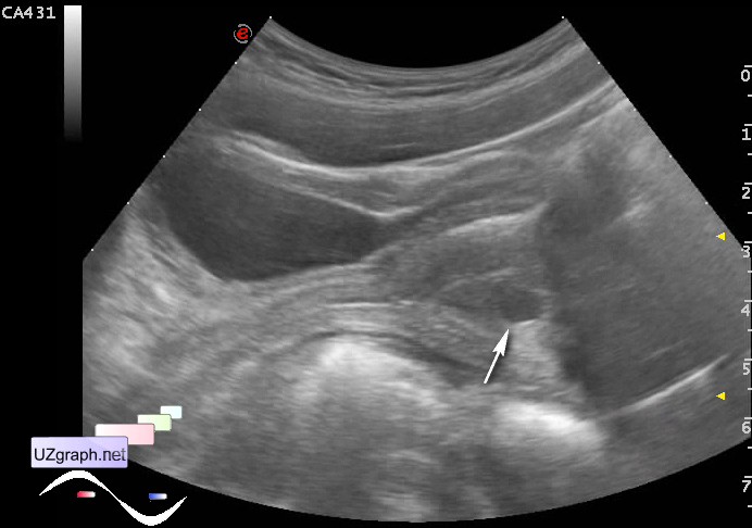
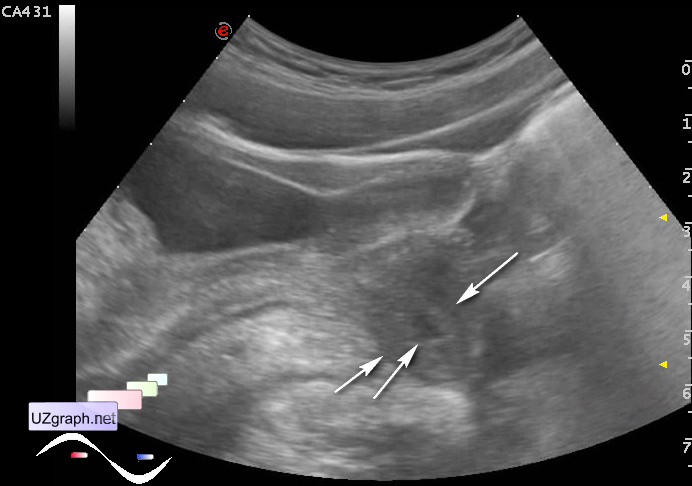
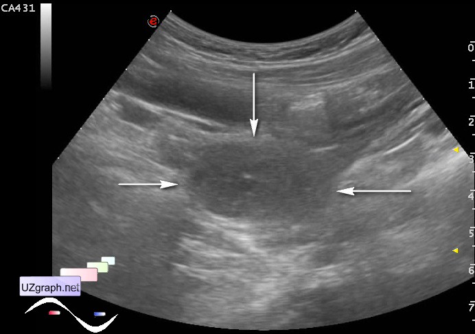
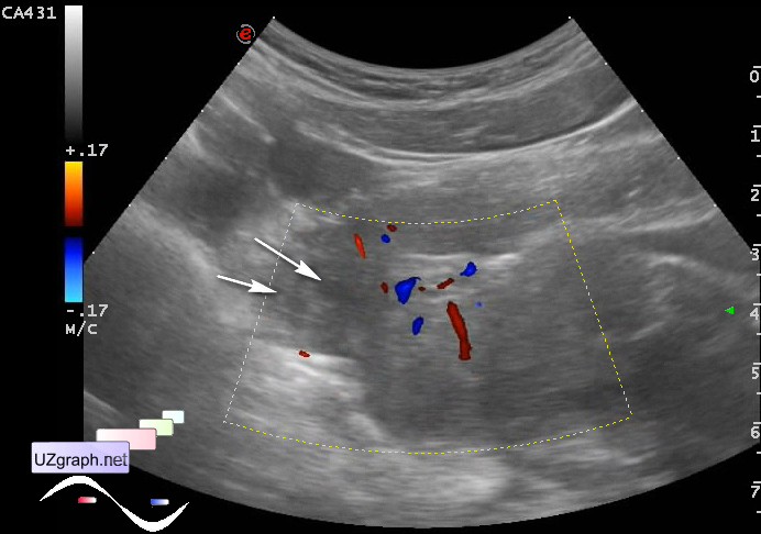
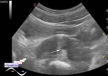
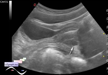
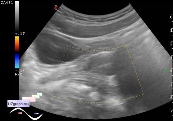
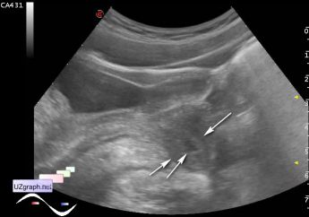
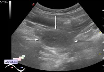
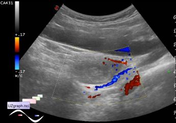
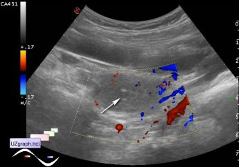
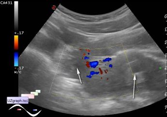
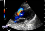
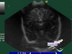
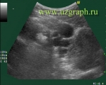
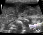
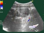
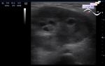
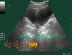
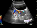
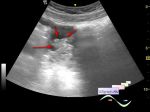
    Girl 13 years old with clinically suspected appendicitis, next day after admission, ultrasound after admission found appendix 6 mm in diameter. Assigned a control ultrasound.

    At ultrasound the appendix is not visualized. The bladder is weak filled, the uterus is located posteriorly, endometrial thickness of 13 mm, in the uterine cavity there is cyst like lesion(attachments 1-3,9), the average diameter of 6.6 mm (early pregnancy? polyp?), Near the uterus formation similar to rudimentary uterine horn(attachment 4) with the oval shaped anechoic lesion(?). Right from the uterus visualized oval shaped heterogeneous structure mass with the small cyst like lesion(attachments 5-9) in the center (ectopic pregnancy? corpus luteum?) at CFM with rich blood supply.

    external link

:: attachments(10) ::

:: file 1 ::

:: file 2 ::

:: file 3 ::

:: file 4 ::

:: file 5 ::

:: file 6 ::

:: file 7 ::

:: file 8 ::

:: file 9 ::

:: file 10 ::


 Random 
Ultrasound cases - Birads 0?
    

Ultrasound cases - Cyst of the base of the brain
    

Ultrasound cases - Cyst but where?
    

Ultrasound cases - Position anomalies of the spleen and left kidney
    

Ultrasound cases - "Ball" in the abdomen
    

 Dictionary 
PASP

PRF

M-mode

LVESV

MC

Qp-Qs

LVEDV

DPD

AC

Freeze


 Calculators 
Type of an infant hip joint

Volume (ellipse formula)

Teicholz systolic cardiac function

Myocardial mass

Pulmonary artery pressure


 Similar topics 
Ultrasound cases - Nucks cyst
    

Ultrasound cases - Hydrosalpinx (saktosalpinx)
    

Ultrasound cases - Portal vein air in the newborn
    

Ultrasound cases - Ovarian teratoma
    

Ultrasound cases - Inguinal hernia with an ovary
    

Ultrasound cases - Giant cyst of the right lateral canal
    

Ultrasound cases - Right ovary changes
    

Ultrasound cases - Ovarian apoplexy 3
    

Ultrasound cases - Hemorrhagic ovarian cyst
    

Ultrasound cases - Patchy uterus