Log in    RU  


Main page :: Forum :: Ultrasound cases - Sonograms, cine-loops etc. ::

Good vs Evil - femur


Tags: Soft tissues sonography, Images, Video, Clinical report, Esaote MyLab 70, Pediatric

   Posts
 
  Good vs Evil - femur
#1

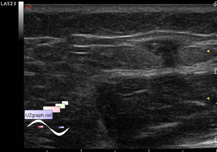
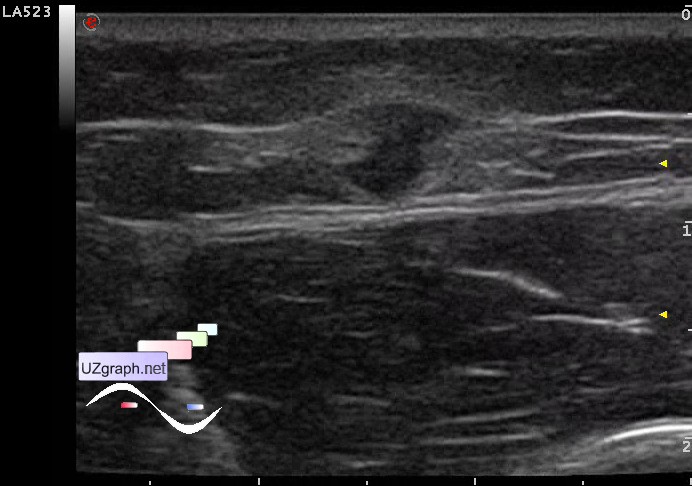
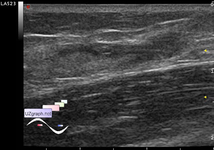
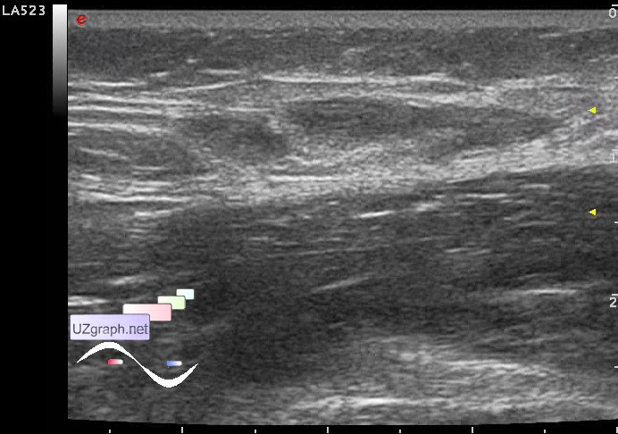
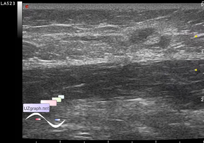
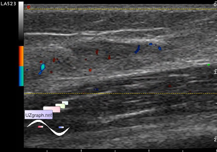
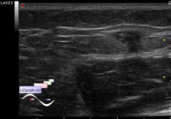
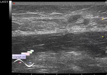
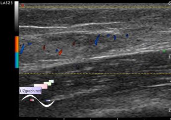
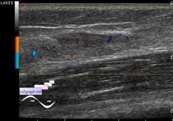
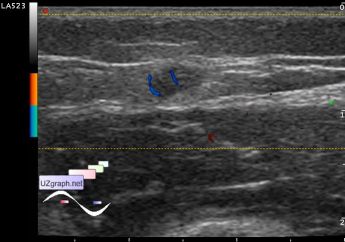
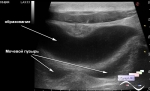
    Child 3 years-old with complaints about the mass of the outer surface of the upper third of femur founded a few days ago accidentally because the child began to itch there, injury or anything else explaining the nature of mass denied.

    Visually, in this field there is a spot of pigmentation to 5 mm, at palpation determined the area of tissues consolidation by the type of tendon running in lengthwise axis of the body, painless on palpation.

    On US visualized heteroechogenic lesion, predominantly hypoechoic with hyperechoic microlesions (microcalcifications?) and hyperechoic area around (infiltration?), with irregular shape and border, at DPD richly blood supplied, 2.5x0.7 cm in size(cr?)

    Recommended consultation of the surgeon, oncologist.

    external link

:: attachments(9) ::

:: file 1 ::

:: file 2 ::

:: file 3 ::

:: file 4 ::

:: file 5 ::

:: file 6 ::

:: file 7 ::

:: file 8 ::

:: file 9 ::

  18:25 23-08-2021
 
#2

    1) myxoma - external link

    2) Desmoid tumor or benign fibromatosis - external link

    3) Metastasis of a renal cell carcinoma in the dorsal aspect of the shoulder - external link

    4) Basal cell carcinoma - external link

    5) Liposarcoma- external link


 Random 
Ultrasound cases - Regional cervical lymphadenitis
    

Ultrasound cases - Non visualization of testicles
    

Ultrasound cases - Neonatal testicular torsion
    

Ultrasound cases - Intestinal intussusception
    

Ultrasound cases - Subcutaneous shoulder lesion
    

 Dictionary 
PADP

HR

FL

Qp-Qs

VTI

PSV

ESD

SV

Doppler angle

LAP


 Calculators 
Teicholz systolic cardiac function

Myocardial mass

Pulmonary artery pressure

Thyroid volume

Estimated fetal weight by Ultrasound


 Similar topics 
Ultrasound cases - Penetrating injury of the femur
    

Ultrasound cases - Cephalohematoma
    

Ultrasound cases - Urachus fistula
    

Ultrasound cases - Cranial mass
    

Ultrasound cases - Pilonidal coccyx cyst
    

Ultrasound cases - Palm fistula, in search of pencil lead
    

Ultrasound cases - Postoperative abdominal wall fistula
    

Ultrasound cases - Nucks cyst
    

Ultrasound cases - Paraproctitis
    

Ultrasound cases - Submandibular area lesion