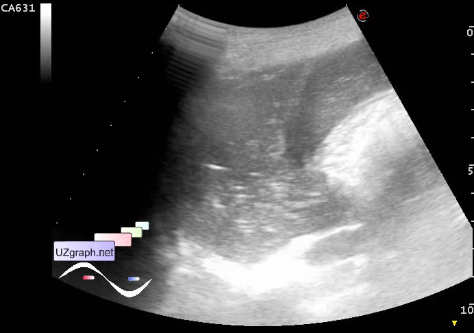
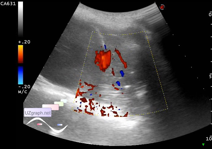
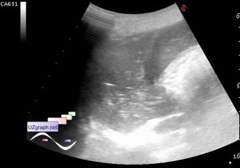
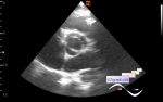
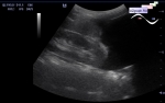
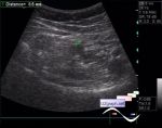
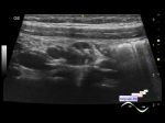
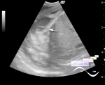
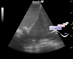
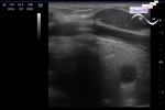
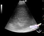
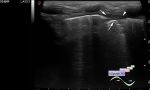
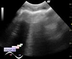
Log in    RU  


Main page :: Forum :: Ultrasound cases - Sonograms, cine-loops etc. ::

Pneumonia


Tags: Chest sonography, Esaote MyLab 70, Images, Video, Clinical report, Pediatric

   Posts
 
  Pneumonia
#1

    A child of 8 years old with a diagnosis of pneumonia, after X-rays sent to the US of the pleural cavity.

    At US from the left side intercostal view visualized portion of airless lung with blood flow at CFM, up to approximately 8 x 6 cm (pneumonia? etc.?)

    external link

:: attachments(3) ::

:: file 1 ::

:: file 2 ::

:: file 3 ::


 Random 
Ultrasound cases - Little Baker cyst
    

Scientific - Playlist Lung ultrasound

Ultrasound cases - Lower abdomen pain 2
    

Ultrasound cases - Liver pseudotumor
    

Admin room - International day of radiology

 Dictionary 
TV

HC

RVOT

EDV

LVESV

EFW

PASP

Gain

CO

TAMEAN


 Calculators 
Resistive index

Pulmonary artery pressure

Type of an infant hip joint

Volume (ellipse formula)

Pressure gradient


 Similar topics 
Ultrasound cases - Chylopericardium, lung atelectasis, cardiomegaly, ascites
    

Ultrasound cases - Diaphragmatic hernia, condition after surgery
    

Ultrasound cases - Hydrothorax
    

Ultrasound cases - One and a half kilograms of happiness
    

Ultrasound cases - Mirror artifact vs collapsed lung
    

Ultrasound cases - Persistent thymus or not
    

Ultrasound cases - Air bronchogram
    

Ultrasound cases - Postoperative hydrothorax with sludge
    

Ultrasound cases - Pneumonia and hydrothorax on the left
    

Ultrasound cases - Diaphragmatic hernia, ectopic spleen & air...