Log in    RU  


Main page :: Forum :: Ultrasound cases - Sonograms, cine-loops etc. ::

Lesion in a female pelvis


Tags: Female pelvis sonography, Medison Sonoace R7, Images, Video, Clinical report, Pediatric

   Posts
 
  Lesion in a female pelvis
#1

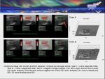
    The child 16 years old from a gynecologist with complaints of a menstrual cycle disorder, by the words before there was an ovarian cyst (there is no evidence of previous ultrasound data)

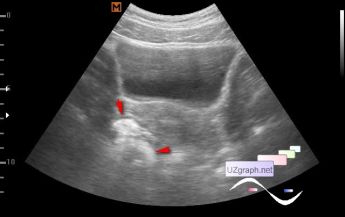
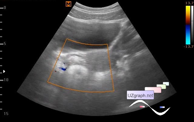
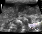
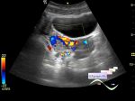
    The left ovary of the usual small-follicular structure, in the projection of the right ovary visualized the lesion of a complex echostructure (the intestine? else?), approximately 50x36mm (anteroposterior dimension can not be obtained - the posterior contour is blurred).

    It is recommended to repeat ultrasound in a week (to exclude the intestines and hidden somewhere ovary).

:: attachments(4) ::

:: file 1 ::

:: file 2 ::

:: file 3 ::

:: file 4 ::

  00:49 02-01-2018
  Donut
#2

    Follow-up ultrasound after 12 days:

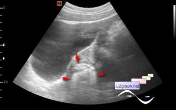
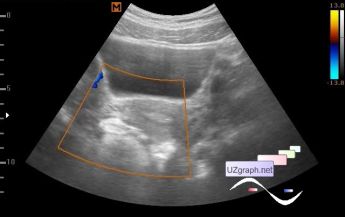
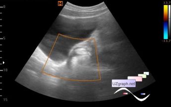
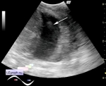
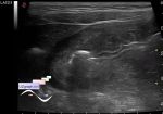
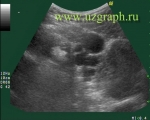
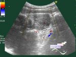
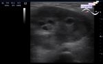
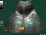
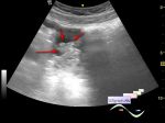
    left ovary 4x2 cm with dominant follicle up to 12 mm, in the projection of the right ovary the lesion of a complex echostructure is visualized - hypoechoic with 2 hyperechogenous inclusions of the oval shape, lesser anteriorly, larger posteriorly resembling a donut structure, a hyperechoic ring from the outside, and in the center a hypoechoic inclusion of the oval shape, the total size of lesion is about 4,5x3x4,5 cm, the blood flow is not visualized at CFM. In comparison with the data of the previous ultrasound without significant changes (teratoma / teratomas of the right ovary?).

    The gynecologist decided to try medicamentous treatment and appointed a repeat ultrasound in 1 month.

    external link

:: attachments(5) ::

:: file 1 ::

:: file 2 ::

:: file 3 ::

:: file 4 ::

:: file 5 ::


 Random 
Ultrasound cases - Lower abdomen pain 2
    

Ultrasound cases - Actualization of the ASA
    

Admin room - Doorways, Goodbye!

Ultrasound cases - Mickey Mouse sign
    

Ultrasound cases - Pelvic dystopia of the hypoplastic kidney
    

 Dictionary 
Color M-mode

VTI

PRF

RVOT

LVESD

TI-RADS

LVEDD

CO

TVUS

PG


 Calculators 
Body surface area

Type of an infant hip joint

Qp:Qs

Teicholz systolic cardiac function

TI-RADS


 Similar topics 
Ultrasound cases - Nucks cyst
    

Ultrasound cases - Hydrosalpinx (saktosalpinx)
    

Ultrasound cases - Portal vein air in the newborn
    

Ultrasound cases - Ovarian teratoma
    

Ultrasound cases - Inguinal hernia with an ovary
    

Ultrasound cases - Giant cyst of the right lateral canal
    

Ultrasound cases - Right ovary changes
    

Ultrasound cases - Ovarian apoplexy 3
    

Ultrasound cases - Hemorrhagic ovarian cyst
    

Ultrasound cases - Patchy uterus