Log in    RU  


Main page :: Forum :: Ultrasound cases - Sonograms, cine-loops etc. ::

2 plus 2


Tags: Abdomen sonography, Thyroid gland sonography, GE Vivid 3, Images, Video, Clinical report, Adult

   Posts
 
  2 plus 2
#1

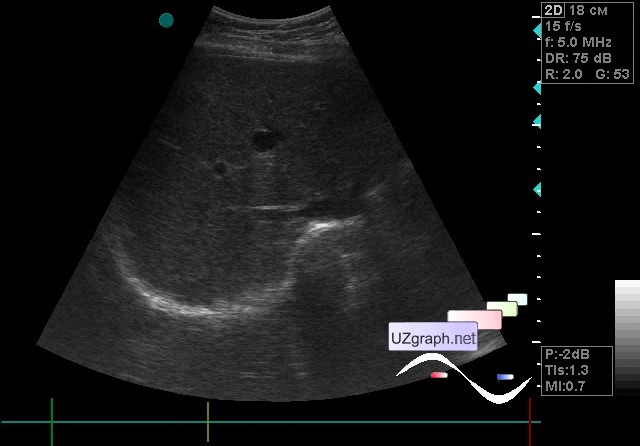
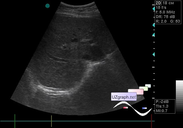
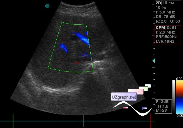
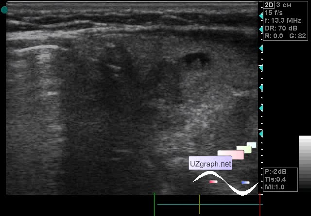
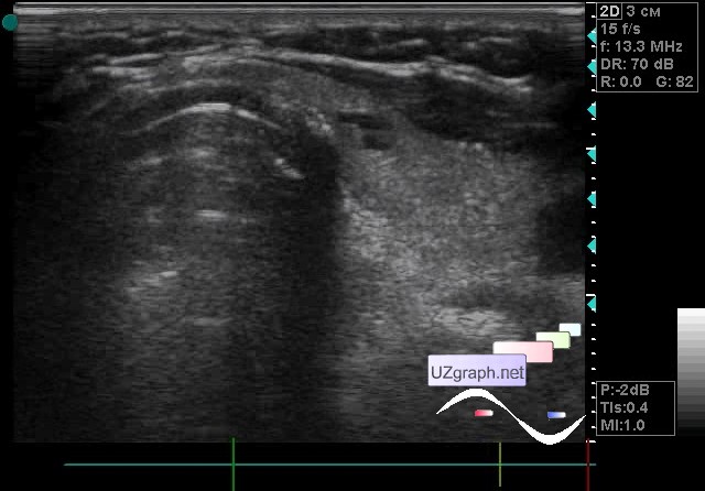
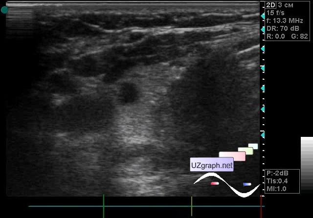
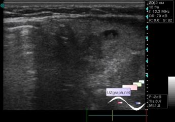
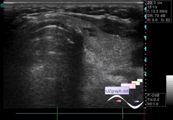
    A 65-year-old patient came for an ultrasound scan of the abdomen and thyroid gland.

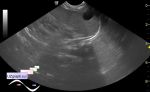
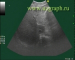
    At ultrasound of the abdomen in the 4th segment of the liver are visualized two cysts.

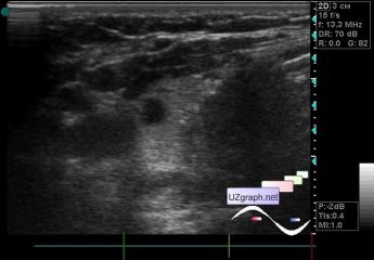
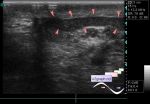
    At ultrasound of the thyroid gland - two nodes, one of the nodes with a near-wall component and a micro hyperechoic inclusion. On the CFM without blood flow (video loops with CFM were lost)

:: attachments(8) ::

:: file 1 ::

:: file 2 ::

:: file 3 ::

:: file 4 ::

:: file 5 ::

:: file 6 ::

:: file 7 ::

:: file 8 ::


 Random 
Ultrasound cases - Interventricular septum defect
    

Ultrasound cases - Acute appendicitis, typhlitis
    

Ultrasound cases - Postoperative hydrothorax with sludge
    

Ultrasound cases - Another PDA
    

Ultrasound cases - Varicose and Baker's cyst
    

 Dictionary 
LVPWd

B-mode

BSA

PSV

GA

BPD

IVSd

FVI

Color M-mode

LN


 Calculators 
Pressure gradient

Teicholz systolic cardiac function

Resistive index

Gestational age by Ultrasound

Body surface area


 Similar topics 
Ultrasound cases - Portal hypertension
    

Ultrasound cases - Lesion in the projection of the adrenal gland 2
    

Ultrasound cases - Gallbladder, saw sign
    

Ultrasound cases - Ileotiphlitis and mesadenitis
    

Ultrasound cases - Cholelithiasis and hyperechoic gallbladder wall
    

Ultrasound cases - Portal vein spontaneous enhancement (mononucleosis)
    

Ultrasound cases - Hypoechoic spleen lesion
    

Ultrasound cases - Acute Appendicitis, periappendicular infiltrate
    

Ultrasound cases - Cavernous Transformation of the Portal Vein
    

Ultrasound cases - Diffuse-focal changes in the liver, thick left kidney