Log in    RU  


Main page :: Forum :: Ultrasound cases - Sonograms, cine-loops etc. ::

I'm normal, it's all the rest ...


Tags: Abdomen sonography, GE Vivid 3, Images, Video, Clinical report, Adult

   Posts
 
  I'm normal, it's all the re...
#1

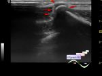
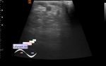
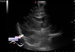
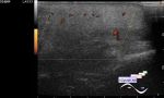
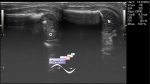
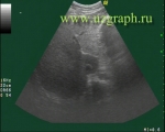
    A 69-year-old patient came for an abdomen ultrasound after gastroscopy, at which a gastric polyps were detected.

    On ultrasound in the projection of 4th segment of the liver a hypoechoic lesion of an irregular shape with an uneven contour is visualized, on the CFM with blood flow, up to 2 cm in size (fat-free zone or focal fatty sparing or, another words, area of normal liver at the background of steatosis / fatty infiltration of the rest liver).

    PS. This localization (segment 4) is typical, which may be associated with the characteristics of the blood supply in this area.

    Also read:

    external link

    external link

    Previously published example:

    https://m.en.uzgraph.ru/forum/...

    external link

:: attachments(2) ::

:: file 1 ::

:: file 2 ::


 Random 
Ultrasound cases - Infiltrate/ phlegmon of the wrist
    

Ultrasound cases - Thyroid lesions 4
    

Admin room - New flash-components

Ultrasound cases - Accessory thymic lobe
    

Ultrasound cases - Postinjection mass
    

 Dictionary 
IVSd

VTI

SF

AC

FVI

LVEDD

TAV

CRL

LMP

ESD


 Calculators 
Resistive index

Pressure gradient

Pulmonary artery pressure

Gestational age by Ultrasound

Myocardial mass


 Similar topics 
Ultrasound cases - Ileal perforation, abscess
    

Ultrasound cases - Portal hypertension
    

Ultrasound cases - Lesion in the projection of the adrenal gland 2
    

Ultrasound cases - Gallbladder, saw sign
    

Ultrasound cases - Ileotiphlitis and mesadenitis
    

Ultrasound cases - Cholelithiasis and hyperechoic gallbladder wall
    

Ultrasound cases - Portal vein spontaneous enhancement (mononucleosis)
    

Ultrasound cases - Hypoechoic spleen lesion
    

Ultrasound cases - Acute Appendicitis, periappendicular infiltrate
    

Ultrasound cases - Cavernous Transformation of the Portal Vein