Log in    RU  


Main page :: Forum :: Ultrasound cases - Sonograms, cine-loops etc. ::

Cystic-solid volume of the thyroid gland


Tags: Thyroid gland sonography, GE Vivid 3, Images, Video, Clinical report, Adult

   Posts
 
  Cystic-solid volume of the thyroid ...
#1

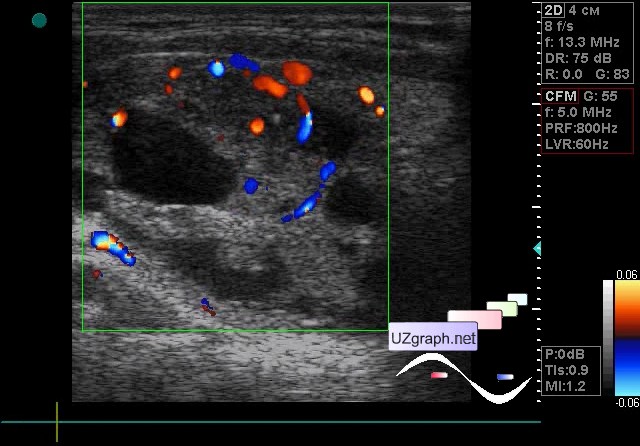
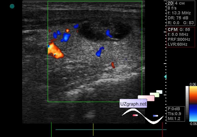
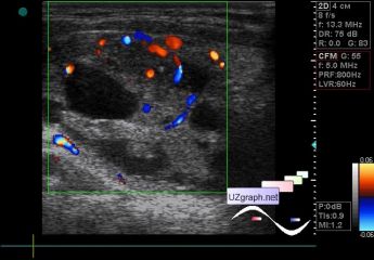
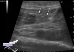
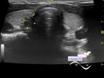
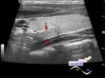
    The patient 18 years old, came to control thyroid ultrasound, thyroid nodules were previously detected at ultrasound, as I understand, a puncture was performed with an unknown result, but there was no surgery after a puncture.

    On the current ultrasound, almost the entire right lobe is occupied by the lesion of a cystic-solid type, with blood flow at the CFM, 4x2x2.5cm.

    A cystic-solid lesion is visualized on the border of the isthmus and the left lobe, with a blood flow at CFM, approximately 0.7x0.7x1.6 cm.

    Recommended puncture and consultation of an endocrinologist.

    external link

:: attachments(3) ::

:: file 1 ::

:: file 2 ::

:: file 3 ::


 Random 
Ultrasound cases - Lesion in a female pelvis
    

Ultrasound cases - Kidney cysts
    

Ultrasound cases - Rectus abdominis muscle contusion
    

Scientific - Varicocele in a child sonography
    

Ultrasound cases - Acute epididymitis
    

 Ad 


 Dictionary 
PADP

PW

RVOT

GS

AC

CO

VTI

LN

SF

EDV


 Calculators 
Pressure gradient

Pulmonary artery pressure

Type of an infant hip joint

Myocardial mass

Gestational age by Ultrasound


 Similar topics 
Ultrasound cases - Tirads 5 multinodular
    

Ultrasound cases - Pediatric Tirads 5
    

Ultrasound cases - Tirads 5
    

Ultrasound cases - Thyroiditis 2
    

Ultrasound cases - Diffuse-focal thyroid changes, parathyroid glands adenomas
    

Ultrasound cases - Accessory Thyroid Lobe
    

Ultrasound cases - Area with hyperechoic dot inclusions
    

Ultrasound cases - Thyroiditis
    

Ultrasound cases - Hypoplasia of the left thyroid lobe
    

Ultrasound cases - Hyperparathyroidism?