Log in    RU  


Main page :: Forum :: Ultrasound cases - Sonograms, cine-loops etc. ::

Appendicolith


Tags: Abdomen sonography, Gastrointestinal sonography, Esaote MyLab 70, Clinical report, Pediatric

   Posts
 
  Appendicolith
#1

    The teenager came to the emergency department of the Children's City Clinical Hospital with suspicion of appendicitis by the ambulance, and was urgently sent for an ultrasound scan.

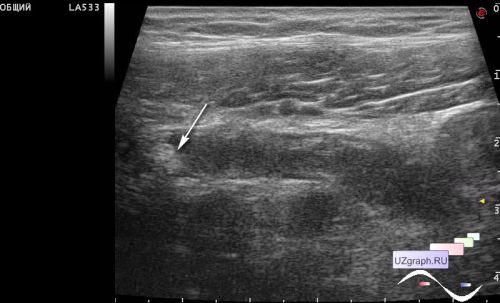
    On ultrasound, a tubular structure 7-9 mm in diameter is visualized in the right iliac region, with an echogenic component at the base (appendicolitis is a stone obstructing the appendix), moderately compressed by the probe.


 Random 
Ultrasound cases - Abdominal syndrome, two in one
    

Ultrasound cases - Cholelithiasis plus 2
    

Ultrasound cases - Lesion in the projection of the pancreas body
    

Ultrasound cases - Muscular IVSD
    

Ultrasound cases - Is it safe to puncture?
    

 Dictionary 
MSD

CFM

RVOT

BSA

M-mode

Aliasing doppler artifact

PSV

PADP

VTI

PW


 Calculators 
Resistive index

Type of an infant hip joint

Body surface area

Thyroid volume

Estimated fetal weight by Ultrasound


 Similar topics 
Ultrasound cases - Hirschsprung disease on ultrasound
    

Ultrasound cases - Intestinal Infiltrate
    

Ultrasound cases - Abdominal cryptorchidism
    

Ultrasound cases - Diffuse heterogeneous liver
    

Ultrasound cases - Postoperative abdominal wall fistula
    

Ultrasound cases - Stump appendicitis
    

Ultrasound cases - Liver scar after resection
    

Ultrasound cases - Subdiaphragmatic movable fluid collection (UFO)
    

Ultrasound cases - Lesion of the spleen or the case when it is easy to not notice
    

Ultrasound cases - I’m normal but others ...