Log in    RU  


Main page :: Forum :: Ultrasound cases - Sonograms, cine-loops etc. ::

Choledochal cyst


Tags: Abdomen sonography, Images, Video, Clinical report, Esaote MyLab 70, Pediatric

   Posts
 
  Choledochal cyst
#1

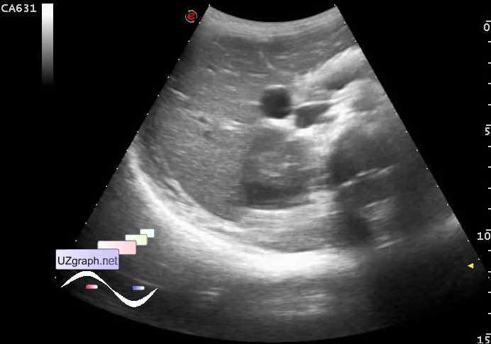
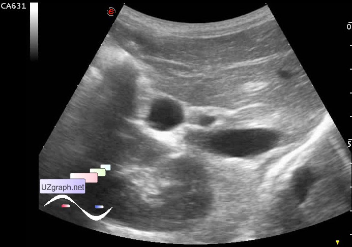
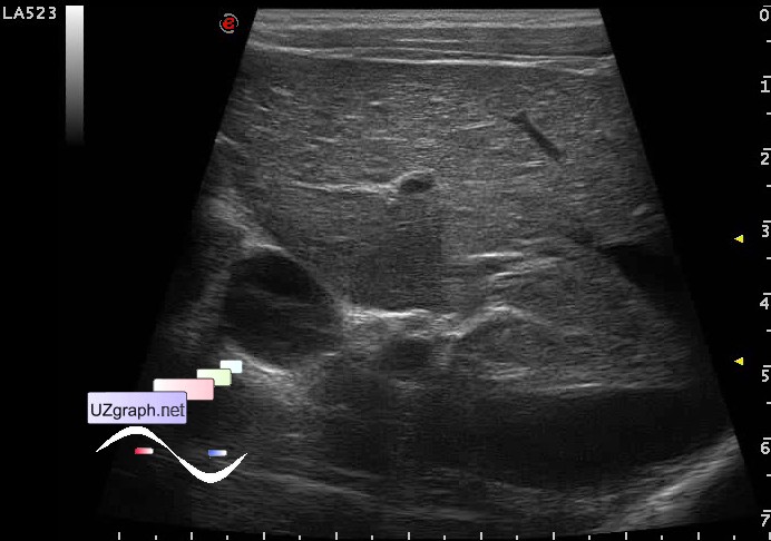
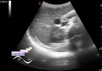
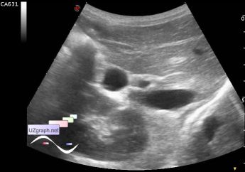
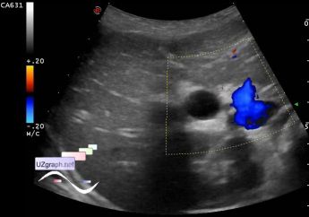
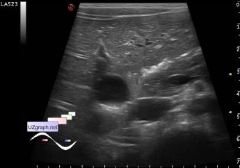
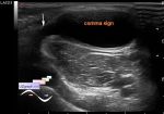
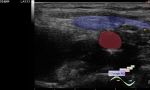
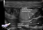
    Child 8 year-old aimed for an abdominal ultrasound because of mesadenitis previously identified in some Moscow child hospital, also in the US from this child hospital described normal oval-shaped GB, at the current ultrasound gallbladder is not visible, but instead cyst, no blood flow at CFM (choledochal cysts?).

    external link

:: attachments(7) ::

:: file 1 ::

:: file 2 ::

:: file 3 ::

:: file 4 ::

:: file 5 ::

:: file 6 ::

:: file 7 ::


 Random 
Ultrasound cases - Left & right ventricular non-compaction
    

Ultrasound cases - Neonatal breast enlargement
    

Ultrasound cases - Chiari net
    

Ultrasound cases - Adolescent Gynecomastia
    

Ultrasound cases - Good vs Evil - thyroid 3
    

 Dictionary 
Color M-mode

LVESD

PG

Freeze

EDV

ESD

IVSd

CRL

RAP

PRF


 Calculators 
Type of an infant hip joint

Body surface area

Myocardial mass

Teicholz systolic cardiac function

Qp:Qs


 Similar topics 
Ultrasound cases - Hirschsprung disease on ultrasound
    

Ultrasound cases - Intestinal Infiltrate
    

Ultrasound cases - Abdominal cryptorchidism
    

Ultrasound cases - Diffuse heterogeneous liver
    

Ultrasound cases - Postoperative abdominal wall fistula
    

Ultrasound cases - Stump appendicitis
    

Ultrasound cases - Liver scar after resection
    

Ultrasound cases - Subdiaphragmatic movable fluid collection (UFO)
    

Ultrasound cases - Lesion of the spleen or the case when it is easy to not notice
    

Ultrasound cases - I’m normal but others ...