Log in    RU  


Main page :: Forum :: Ultrasound cases - Sonograms, cine-loops etc. ::

Such different ovaries


Tags: Female pelvis sonography, Medison Sonoace R7, Images, Video, Clinical report, Pediatric

   Posts
 
  Such different ovaries
#1

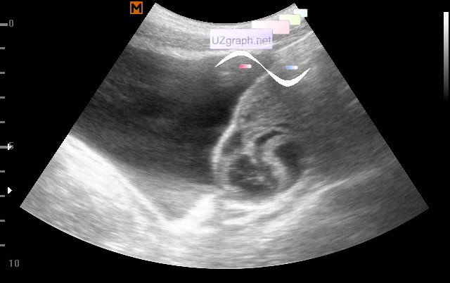
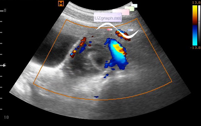
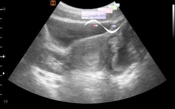
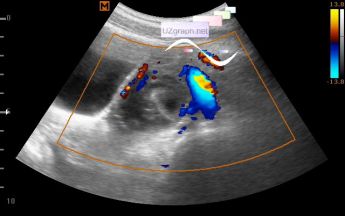
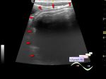
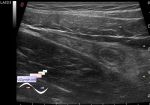
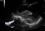
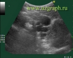
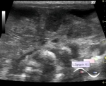
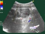
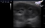
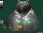
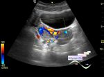
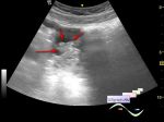
    A teenager of 17 years, with complaints about a disturbance of the MC.

    The right ovary of the small-follicular structure, with the follicles located mainly along the periphery, with a thickened stroma of increased echogenicity (according to PCOS type), of normal size; Left approximately up to 4.5 cm in size, of a complex structure, including a hemorrhagic cyst-type component, as well as cysts with a thick hyperechogenic wall, a solid component (teratoma / dermoid? hemorrhagic cyst? etc.).

    external link

:: attachments(4) ::

:: file 1 ::

:: file 2 ::

:: file 3 ::

:: file 4 ::

  14:34 27-07-2017
 
#2

    The gynecologist prescribed to the girl a Duphaston, the monthly came, at the control female pelvis TA ultrasound - everything is fine.

    PS. So it was a such hemorrhagic cyst.


 Random 
Ultrasound cases - Acute Appendicitis
    

Ultrasound cases - Condition after surgery - groin
    

Ultrasound cases - Testicular torsion after surgery and thyroid
    

Ultrasound cases - Thickening of the duodenum wall
    

Ultrasound cases - Traumatic epididymitis
    

 Dictionary 
FNA

CFM

TAV

PADP

ESV

TI-RADS

DPD

TV

LVPWd

EDD


 Calculators 
TI-RADS

Type of an infant hip joint

Resistive index

Volume (ellipse formula)

Thyroid volume


 Similar topics 
Ultrasound cases - Right ovary changes
    

Ultrasound cases - Ovarian apoplexy 3
    

Ultrasound cases - Hemorrhagic ovarian cyst
    

Ultrasound cases - Patchy uterus
    

Ultrasound cases - Fibroid and nabothian cyst
    

Ultrasound cases - Small pelvis lesion, presumably hydrocolpos
    

Ultrasound cases - Something in the female pelvis
    

Ultrasound cases - Uterine myoma(fibroid)
    

Ultrasound cases - Giant mass in the female pelvis
    

Ultrasound cases - Gallbladder, free fluid, etc.