Log in    RU  


Main page :: Forum :: Ultrasound cases - Sonograms, cine-loops etc. ::

Something in the female pelvis


Tags: Female pelvis sonography, GE Vivid 3, Images, Video, Clinical report, Adult

   Posts
 
  Something in the female pelvis
#1

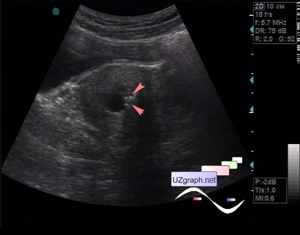
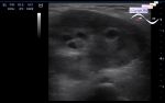
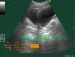
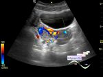
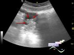
    A 38-year-old female patient came to the university public clinic for the abdomen ultrasound.

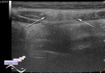
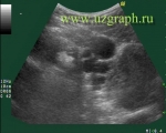
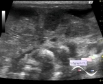
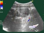
    On abdomen ultrasound without features, in a small pelvis in the projection of the uterus cervix, more than 5 cysts (nabothian) up to 9 mm are visualized, next to one of the cysts, an area with hyperechoic microinclusions (?) is visualized.

    Was recommended to monitor the ultrasound of the female pelvis after filling the bladder or TVUS.

    external link

:: attachments(2) ::

:: file 1 ::

:: file 2 ::

  09:34 26-09-2018
 
#2

    Endometrial and Endocervical Micro Echogenic Foci

    external link

    "Clinical follow-up in 62 patients showed that this condition was mostly benign (endometrioid carcinoma developed in 1 patient)."

  12:35 09-08-2020
 
#3

    At Facebook added to the differential diagnosis a versions of an old abortion and a calcified polyp.


 Random 
Ultrasound cases - Simple splenic cyst
    

Ultrasound cases - Patent ductus arteriosus(PDA)
    

Ultrasound cases - Penetrating wound of the submandibular region
    

Ultrasound cases - Scrotum hematocele
    

Ultrasound cases - Phantom testicle
    

 Dictionary 
TI-RADS

EDD

BPD

ESV

PA

GS

EDV

PG

PADP

LVESD


 Calculators 
Pulmonary artery pressure

Volume (ellipse formula)

Estimated fetal weight by Ultrasound

Myocardial mass

Type of an infant hip joint


 Similar topics 
Ultrasound cases - Nucks cyst
    

Ultrasound cases - Hydrosalpinx (saktosalpinx)
    

Ultrasound cases - Portal vein air in the newborn
    

Ultrasound cases - Ovarian teratoma
    

Ultrasound cases - Inguinal hernia with an ovary
    

Ultrasound cases - Giant cyst of the right lateral canal
    

Ultrasound cases - Right ovary changes
    

Ultrasound cases - Ovarian apoplexy 3
    

Ultrasound cases - Hemorrhagic ovarian cyst
    

Ultrasound cases - Patchy uterus