Log in    RU  


Main page :: Forum :: Ultrasound cases - Sonograms, cine-loops etc. ::

Hypoechoic node


Tags: Thyroid gland sonography, Images, Video, Clinical report, Esaote MyLab 70, Pediatric

   Posts
 
  Hypoechoic node
#1

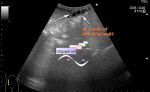
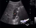
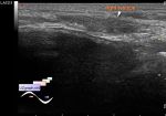
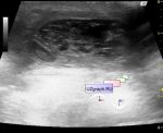
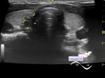
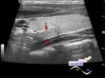
    Teenager on school medical examination by schedule(without complaints), unremarkable by the conclusion of the endocrinologist.

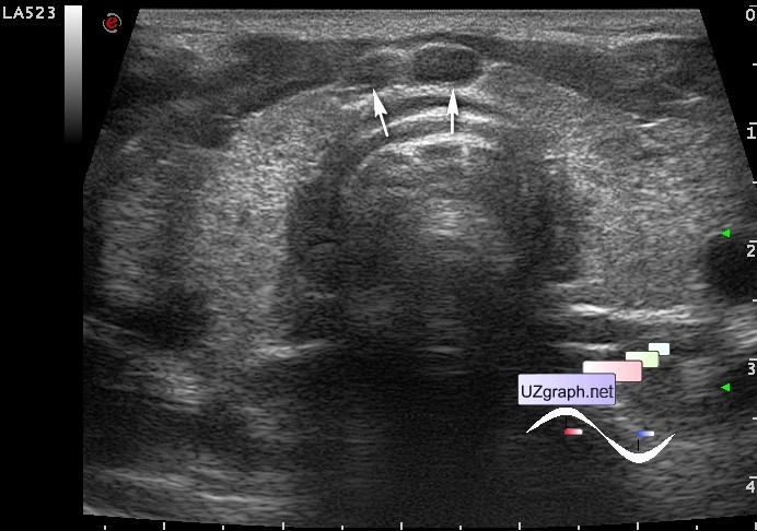
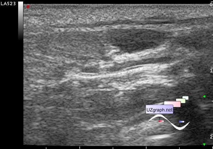
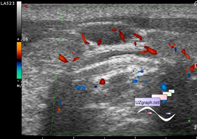
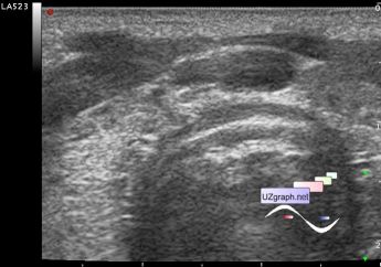
    At ultrasound thyroid gland volume about 15 ml with diffusely inhomogeneous structure and increased blood flow in the CFM (Hashimoto' s?). In the isthmus there are 1-2 an / hypoechoic lesions, oval shape, with a clear outline, at CFM is well supplied with blood (adenomatous hyperplasia? LN?).

:: attachments(5) ::

:: file 1 ::

:: file 2 ::

:: file 3 ::

:: file 4 ::

:: file 5 ::

  16:08 20-10-2018
 
#2

    With further analysis, I came to the conclusion that these are pre-tracheal lymph nodes.


 Random 
Ultrasound cases - Simple splenic cyst
    

Ultrasound cases - Prepatellar bursitis
    

Ultrasound cases - Chiari net
    

Ultrasound cases - Effusion in the pericardial cavity
    

Ultrasound cases - Exclude bleeding
    

 Dictionary 
PA

Color M-mode

LMP

PW

CW

BSA

LVOT

EF

Doppler angle

TVUS


 Calculators 
Pressure gradient

Gestational age by Ultrasound

Body surface area

Teicholz systolic cardiac function

Resistive index


 Similar topics 
Ultrasound cases - Tirads 5 multinodular
    

Ultrasound cases - Pediatric Tirads 5
    

Ultrasound cases - Tirads 5
    

Ultrasound cases - Thyroiditis 2
    

Ultrasound cases - Diffuse-focal thyroid changes, parathyroid glands adenomas
    

Ultrasound cases - Accessory Thyroid Lobe
    

Ultrasound cases - Area with hyperechoic dot inclusions
    

Ultrasound cases - Thyroiditis
    

Ultrasound cases - Hypoplasia of the left thyroid lobe
    

Ultrasound cases - Cystic-solid volume of the thyroid gland