Log in    RU  


Main page :: Forum :: Ultrasound cases - Sonograms, cine-loops etc. ::

Good vs Evil - thyroid 3


Tags: Thyroid gland sonography, Soft tissues sonography, Musculoskeletal sonography, Medison Sonoace R7, Images, Video, Clinical report, Pediatric

   Posts
 
  Good vs Evil - thyroid 3
#1

    A child of 15 years old turned to the pediatrician with complaints about episodes of a feeling of cold in hands and weakness. Pediatrician directed child to examination including the endocrinologist. The endocrinologist has appointed the US of thyroid.

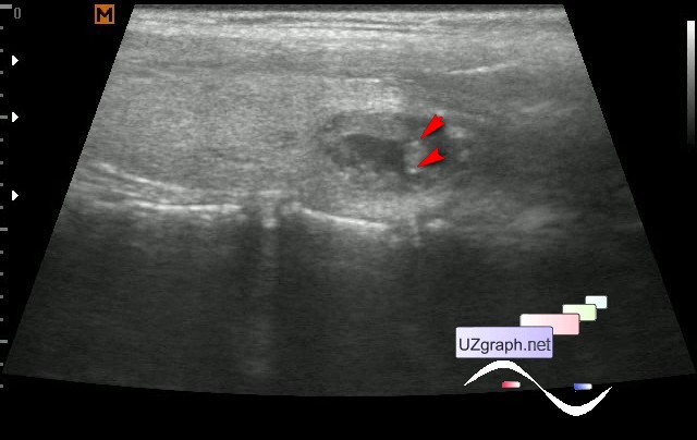
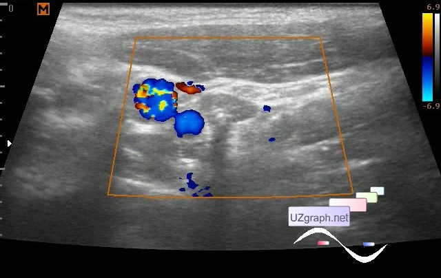
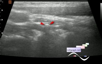
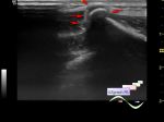
    At ultrasound in the left lobe of the thyroid gland there is a complex cystic lesion with hyperechoic microinclusions giving the artifact of the comet' s tail (microcalcinates?), at CFM is richly supplied by blood, about 13x8 mm in size, with a width greater than height shape (cr?)

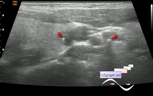
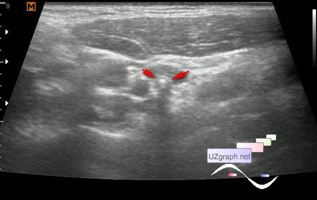
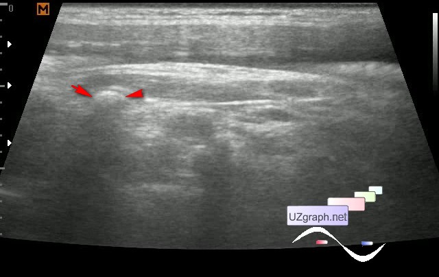
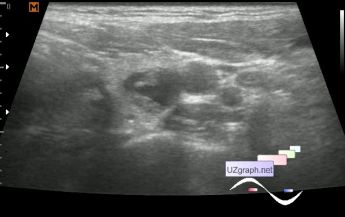
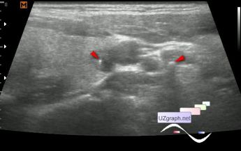
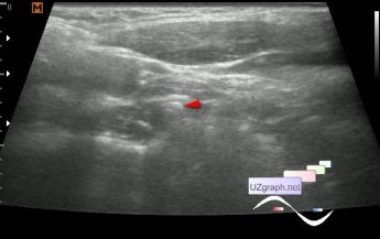
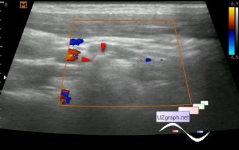
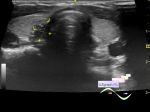
    In one view with the thyroid lesion, lateral to the main vessels of the neck (CCA, IJV), 2 predominantly hyperechogenic lesions up to 4 mm are visualized in the projection of the muscle tissue, giving an acoustic shadow, at CFM without blood flow (mts?)

    The child was directed to an oncologist by cito.

    external link

:: attachments(11) ::

:: file 1 ::

:: file 2 ::

:: file 3 ::

:: file 4 ::

:: file 5 ::

:: file 6 ::

:: file 7 ::

:: file 8 ::

:: file 9 ::

:: file 10 ::

:: file 11 ::

  18:30 10-10-2017
 
#2

    Received the information that in the profile institution was made an ultrasound, they said that most likely the node is benign, but just in case they will make a puncture.

    As for mts, I myself was not sure, maybe that' s just looked like a lesion a part of the transverse process of the vertebra (tuberculum caroticum), but in such cases there is no oversafe.

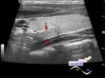
    I understand the logic of colleagues, they notice that this node is not fully solid, but partially solid and cystic, and in such nodes the risk of cancer is much lower, but the suspicion by ultrasound for microcalcinates in any case requires puncture ...

  14:50 17-10-2017
 
#3

    According to the results of the puncture - " not dense colloid. No cells of the epithelium of the thyroid gland were found. "

    Clinical diagnosis: goiter.

  15:40 17-10-2017
 
#4

    A case of papillary microcarcinoma of the thyroid with abundant colloid (masquerading as colloid goiter with papillary hyperplasia)

    external link

    " The coexistence of colloid nodule and thyroid carcinoma was found in 122 cases (38.7%)"

    external link


 Random 
Ultrasound cases - Suspected appendicitis
    

Ultrasound cases - Myxomatous degeneration of MV
    

Ultrasound cases - Thigh lesion
    

Ultrasound cases - Clinically suspected appendicitis
    

Ultrasound cases - Large axillary lesion
    

 Dictionary 
CFM

CW

EDD

Gain

PW

EFW

SF

HR

PG

HC


 Calculators 
Teicholz systolic cardiac function

Gestational age by Ultrasound

TI-RADS

Type of an infant hip joint

Qp:Qs


 Similar topics 
Ultrasound cases - Tirads 5 multinodular
    

Ultrasound cases - Pediatric Tirads 5
    

Ultrasound cases - Tirads 5
    

Ultrasound cases - Thyroiditis 2
    

Ultrasound cases - Diffuse-focal thyroid changes, parathyroid glands adenomas
    

Ultrasound cases - Accessory Thyroid Lobe
    

Ultrasound cases - Area with hyperechoic dot inclusions
    

Ultrasound cases - Thyroiditis
    

Ultrasound cases - Hypoplasia of the left thyroid lobe
    

Ultrasound cases - Cystic-solid volume of the thyroid gland